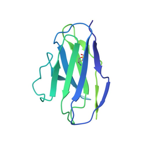
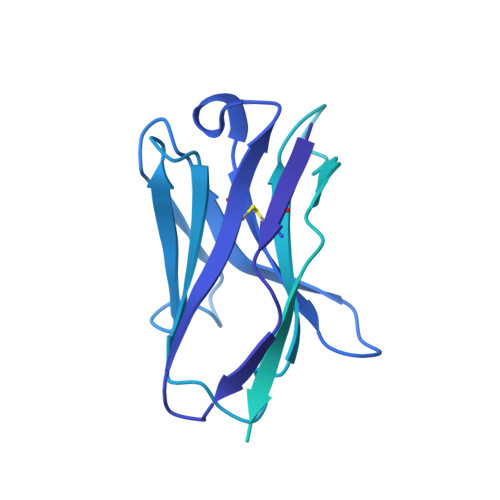
6X3S

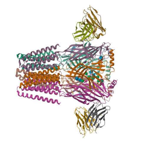
Human GABAA receptor alpha1-beta2-gamma2 subtype in complex with bicuculline methbromide


Experimental Data Snapshot

  • Method: ELECTRON MICROSCOPY
  • Resolution: 3.12 Å
  • Aggregation State: PARTICLE 
  • Reconstruction Method: SINGLE PARTICLE 

wwPDB Validation   3D Report Full Report


This is version 1.3 of the entry. See complete history


Literature

Shared structural mechanisms of general anaesthetics and benzodiazepines.

Kim, J.J.Gharpure, A.Teng, J.Zhuang, Y.Howard, R.J.Zhu, S.Noviello, C.M.Walsh Jr., R.M.Lindahl, E.Hibbs, R.E.

(2020) Nature 585: 303-308

  • DOI: https://doi.org/10.1038/s41586-020-2654-5
  • Primary Citation of Related Structures:  
    6X3S, 6X3T, 6X3U, 6X3V, 6X3W, 6X3X, 6X3Z, 6X40

  • PubMed Abstract: 

    Most general anaesthetics and classical benzodiazepine drugs act through positive modulation of γ-aminobutyric acid type A (GABA A ) receptors to dampen neuronal activity in the brain 1-5 . However, direct structural information on the mechanisms of general anaesthetics at their physiological receptor sites is lacking. Here we present cryo-electron microscopy structures of GABA A receptors bound to intravenous anaesthetics, benzodiazepines and inhibitory modulators. These structures were solved in a lipidic environment and are complemented by electrophysiology and molecular dynamics simulations. Structures of GABA A receptors in complex with the anaesthetics phenobarbital, etomidate and propofol reveal both distinct and common transmembrane binding sites, which are shared in part by the benzodiazepine drug diazepam. Structures in which GABA A receptors are bound by benzodiazepine-site ligands identify an additional membrane binding site for diazepam and suggest an allosteric mechanism for anaesthetic reversal by flumazenil. This study provides a foundation for understanding how pharmacologically diverse and clinically essential drugs act through overlapping and distinct mechanisms to potentiate inhibitory signalling in the brain.


  • Organizational Affiliation

    Department of Neuroscience, University of Texas Southwestern Medical Center, Dallas, TX, USA.


Macromolecules
Find similar proteins by:  (by identity cutoff)  |  3D Structure
Entity ID: 1
MoleculeChains Sequence LengthOrganismDetailsImage
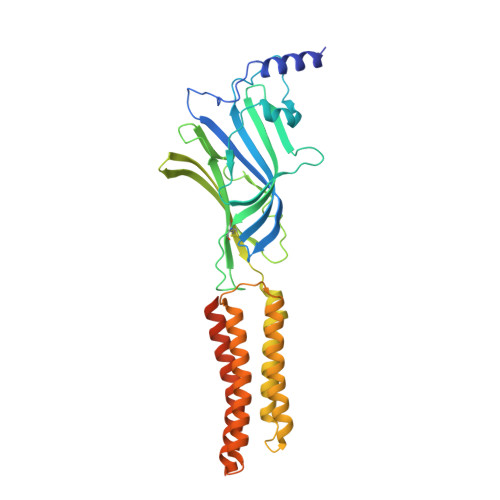
Gamma-aminobutyric acid receptor subunit beta-2
A, C
364Homo sapiensMutation(s): 0 
Gene Names: GABRB2
Membrane Entity: Yes 
UniProt & NIH Common Fund Data Resources
Find proteins for P47870 (Homo sapiens)
Explore P47870 
Go to UniProtKB:  P47870
PHAROS:  P47870
GTEx:  ENSG00000145864 
Entity Groups  
Sequence Clusters30% Identity50% Identity70% Identity90% Identity95% Identity100% Identity
UniProt GroupP47870
Glycosylation
Glycosylation Sites: 2Go to GlyGen: P47870-2
Sequence Annotations
Expand
  • Reference Sequence
Find similar proteins by:  (by identity cutoff)  |  3D Structure
Entity ID: 2
MoleculeChains Sequence LengthOrganismDetailsImage
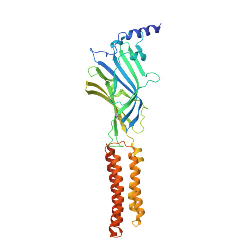
Gamma-aminobutyric acid receptor subunit alpha-1
B, D
358Homo sapiensMutation(s): 0 
Gene Names: GABRA1
Membrane Entity: Yes 
UniProt & NIH Common Fund Data Resources
Find proteins for P14867 (Homo sapiens)
Explore P14867 
Go to UniProtKB:  P14867
PHAROS:  P14867
GTEx:  ENSG00000022355 
Entity Groups  
Sequence Clusters30% Identity50% Identity70% Identity90% Identity95% Identity100% Identity
UniProt GroupP14867
Glycosylation
Glycosylation Sites: 1Go to GlyGen: P14867-1
Sequence Annotations
Expand
  • Reference Sequence
Find similar proteins by:  (by identity cutoff)  |  3D Structure
Entity ID: 3
MoleculeChains Sequence LengthOrganismDetailsImage
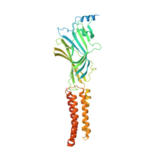
Gamma-aminobutyric acid type A receptor subunit gamma-2417Homo sapiensMutation(s): 0 
Membrane Entity: Yes 
UniProt & NIH Common Fund Data Resources
Find proteins for P18507 (Homo sapiens)
Explore P18507 
Go to UniProtKB:  P18507
PHAROS:  P18507
GTEx:  ENSG00000113327 
Entity Groups  
Sequence Clusters30% Identity50% Identity70% Identity90% Identity95% Identity100% Identity
UniProt GroupP18507
Glycosylation
Glycosylation Sites: 1Go to GlyGen: P18507-2
Sequence Annotations
Expand
  • Reference Sequence
Find similar proteins by:  (by identity cutoff)  |  3D Structure
Entity ID: 4
MoleculeChains Sequence LengthOrganismDetailsImage
Kappa Fab Light ChainF [auth I],
H [auth L]
213Mus musculusMutation(s): 0 
Entity Groups  
Sequence Clusters30% Identity50% Identity70% Identity90% Identity95% Identity100% Identity
Sequence Annotations
Expand
  • Reference Sequence
Find similar proteins by:  (by identity cutoff)  |  3D Structure
Entity ID: 5
MoleculeChains Sequence LengthOrganismDetailsImage
IgG2b Fab Heavy ChainG [auth J],
I [auth K]
454Mus musculusMutation(s): 0 
Entity Groups  
Sequence Clusters30% Identity50% Identity70% Identity90% Identity95% Identity100% Identity
Sequence Annotations
Expand
  • Reference Sequence
Oligosaccharides

Help

Entity ID: 6
MoleculeChains Length2D Diagram Glycosylation3D Interactions
beta-D-mannopyranose-(1-4)-2-acetamido-2-deoxy-beta-D-glucopyranose-(1-4)-2-acetamido-2-deoxy-beta-D-glucopyranoseJ [auth F],
L [auth H]
3N-Glycosylation
Glycosylation Resources
GlyTouCan:  G15407YE
GlyCosmos:  G15407YE
GlyGen:  G15407YE
Entity ID: 7
MoleculeChains Length2D Diagram Glycosylation3D Interactions
alpha-D-mannopyranose-(1-2)-alpha-D-mannopyranose-(1-2)-alpha-D-mannopyranose-(1-3)-[alpha-D-mannopyranose-(1-2)-alpha-D-mannopyranose-(1-6)-alpha-D-mannopyranose-(1-6)]beta-D-mannopyranose-(1-4)-2-acetamido-2-deoxy-beta-D-glucopyranose-(1-4)-2-acetamido-2-deoxy-beta-D-glucopyranoseK [auth G]9N-Glycosylation
Glycosylation Resources
GlyTouCan:  G04414GO
GlyCosmos:  G04414GO
GlyGen:  G04414GO
Entity ID: 8
MoleculeChains Length2D Diagram Glycosylation3D Interactions
2-acetamido-2-deoxy-beta-D-glucopyranose-(1-4)-2-acetamido-2-deoxy-beta-D-glucopyranose
M
2N-Glycosylation
Glycosylation Resources
GlyTouCan:  G42666HT
GlyCosmos:  G42666HT
GlyGen:  G42666HT
Small Molecules
Ligands 3 Unique
IDChains Name / Formula / InChI Key2D Diagram3D Interactions
J94 (Subject of Investigation/LOI)
Query on J94

Download Ideal Coordinates CCD File 
O [auth A],
Q [auth C]
(5S)-6,6-dimethyl-5-[(6R)-8-oxo-6,8-dihydrofuro[3,4-e][1,3]benzodioxol-6-yl]-5,6,7,8-tetrahydro[1,3]dioxolo[4,5-g]isoquinolin-6-ium
C21 H20 N O6
WDIQXKYUSINZME-RBUKOAKNSA-N
NAG
Query on NAG

Download Ideal Coordinates CCD File 
N [auth A],
P [auth C],
R [auth D]
2-acetamido-2-deoxy-beta-D-glucopyranose
C8 H15 N O6
OVRNDRQMDRJTHS-FMDGEEDCSA-N
MAN
Query on MAN

Download Ideal Coordinates CCD File 
S [auth E]alpha-D-mannopyranose
C6 H12 O6
WQZGKKKJIJFFOK-PQMKYFCFSA-N
Experimental Data & Validation

Experimental Data

  • Method: ELECTRON MICROSCOPY
  • Resolution: 3.12 Å
  • Aggregation State: PARTICLE 
  • Reconstruction Method: SINGLE PARTICLE 
EM Software:
TaskSoftware PackageVersion
RECONSTRUCTIONRELION3.0
MODEL REFINEMENTPHENIX

Structure Validation

View Full Validation Report


Entry History & Funding Information

Deposition Data


Funding OrganizationLocationGrant Number
National Institutes of Health/National Institute on Drug Abuse (NIH/NIDA)United StatesDA037492
National Institutes of Health/National Institute on Drug Abuse (NIH/NIDA)United StatesDA042072
National Institutes of Health/National Institute of Neurological Disorders and Stroke (NIH/NINDS)United StatesNS095899
Welch FoundationUnited StatesI-1812
American Heart AssociationUnited States20POST35200127

Revision History  (Full details and data files)

  • Version 1.0: 2020-09-09
    Type: Initial release
  • Version 1.1: 2020-09-16
    Changes: Database references
  • Version 1.2: 2020-09-23
    Changes: Database references
  • Version 1.3: 2024-11-13
    Changes: Data collection, Database references, Structure summary