6X39 | pdb_00006x39

Crystal structure of the FN5 domain of Mouse Lar

  • Classification: CELL ADHESION
  • Organism(s): Mus musculus
  • Expression System: Escherichia coli
  • Mutation(s): No 

  • Deposited: 2020-05-21 Released: 2021-05-26 
  • Deposition Author(s): Bouyain, S., Kawakami, J.E.
  • Funding Organization(s): National Institutes of Health/National Institute of Arthritis and Musculoskeletal and Skin Diseases (NIH/NIAMS)

Experimental Data Snapshot

  • Method: X-RAY DIFFRACTION
  • Resolution: 1.70 Å
  • R-Value Free: 
    0.190 (Depositor), 0.192 (DCC) 
  • R-Value Work: 
    0.164 (Depositor), 0.165 (DCC) 
  • R-Value Observed: 
    0.166 (Depositor) 

Starting Model: experimental
View more details

wwPDB Validation 3D Report Full Report

Validation slider image for 6X39

Ligand Structure Quality Assessment 


This is version 1.2 of the entry. See complete history

Literature

Complex protein interactions mediate Drosophila Lar function in muscle tissue.

Kawakami, J.Brooks, D.Zalmai, R.Hartson, S.D.Bouyain, S.Geisbrecht, E.R.

(2022) PLoS One 17: e0269037-e0269037

  • DOI: https://doi.org/10.1371/journal.pone.0269037
  • Primary Citation Related Structures: 
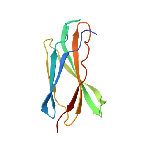
    6X38, 6X39, 6X3A

  • PubMed Abstract: 

    The type IIa family of receptor protein tyrosine phosphatases (RPTPs), including Lar, RPTPσ and RPTPδ, are well-studied in coordinating actin cytoskeletal rearrangements during axon guidance and synaptogenesis. To determine whether this regulation is conserved in other tissues, interdisciplinary approaches were utilized to study Lar-RPTPs in the Drosophila musculature. Here we find that the single fly ortholog, Drosophila Lar (Dlar), is localized to the muscle costamere and that a decrease in Dlar causes aberrant sarcomeric patterning, deficits in larval locomotion, and integrin mislocalization. Sequence analysis uncovered an evolutionarily conserved Lys-Gly-Asp (KGD) signature in the extracellular region of Dlar. Since this tripeptide sequence is similar to the integrin-binding Arg-Gly-Asp (RGD) motif, we tested the hypothesis that Dlar directly interacts with integrin proteins. However, structural analyses of the fibronectin type III domains of Dlar and two vertebrate orthologs that include this conserved motif indicate that this KGD tripeptide is not accessible and thus unlikely to mediate physical interactions with integrins. These results, together with the proteomics identification of basement membrane (BM) proteins as potential ligands for type IIa RPTPs, suggest a complex network of protein interactions in the extracellular space that may mediate Lar function and/or signaling in muscle tissue.


  • Organizational Affiliation
    • Department of Cell and Molecular Biology and Biochemistry, University of Missouri-Kansas City, Kansas City, MO, United States of America.

Macromolecule Content 

  • Total Structure Weight: 11.79 kDa 
  • Atom Count: 888 
  • Modeled Residue Count: 100 
  • Deposited Residue Count: 109 
  • Unique protein chains: 1

Macromolecules

Find similar proteins by:|  3D Structure
Entity ID: 1
MoleculeChains  Sequence LengthOrganismDetailsImage
Receptor-type tyrosine-protein phosphatase F109Mus musculusMutation(s): 0 
Gene Names: PtprfLar
EC: 3.1.3.48
UniProt
Find proteins for A2A8L5 (Mus musculus)
Explore A2A8L5 
Go to UniProtKB:  A2A8L5
Entity Groups
Sequence Clusters30% Identity50% Identity70% Identity90% Identity95% Identity100% Identity
UniProt GroupA2A8L5
Sequence Annotations
Expand
Reference Sequence

Small Molecules

Ligands 1 Unique
IDChains Name / Formula / InChI Key2D Diagram3D Interactions
SOR

Query on SOR


Download:Ideal Coordinates CCD File
B [auth A]sorbitol
C6 H14 O6
FBPFZTCFMRRESA-JGWLITMVSA-N

Experimental Data & Validation

Experimental Data

  • Method: X-RAY DIFFRACTION
  • Resolution: 1.70 Å
  • R-Value Free:  0.190 (Depositor), 0.192 (DCC) 
  • R-Value Work:  0.164 (Depositor), 0.165 (DCC) 
  • R-Value Observed: 0.166 (Depositor) 
Space Group: P 32 2 1
Unit Cell:
Length ( Å )Angle ( ˚ )
a = 86.11α = 90
b = 86.11β = 90
c = 29.344γ = 120
Software Package:
Software NamePurpose
PHENIXphasing
PHENIXrefinement
HKL-2000data reduction
HKL-2000data scaling
SERGUIdata collection

Structure Validation

View Full Validation Report


Ligand Structure Quality Assessment 


Entry History 

& Funding Information

Deposition Data


Funding OrganizationLocationGrant Number
National Institutes of Health/National Institute of Arthritis and Musculoskeletal and Skin Diseases (NIH/NIAMS)United StatesR21AR066264

Revision History  (Full details and data files)

  • Version 1.0: 2021-05-26
    Type: Initial release
  • Version 1.1: 2022-06-22
    Changes: Database references
  • Version 1.2: 2024-04-03
    Changes: Data collection, Refinement description