6X1Y | pdb_00006x1y

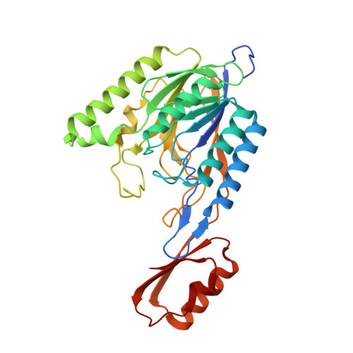
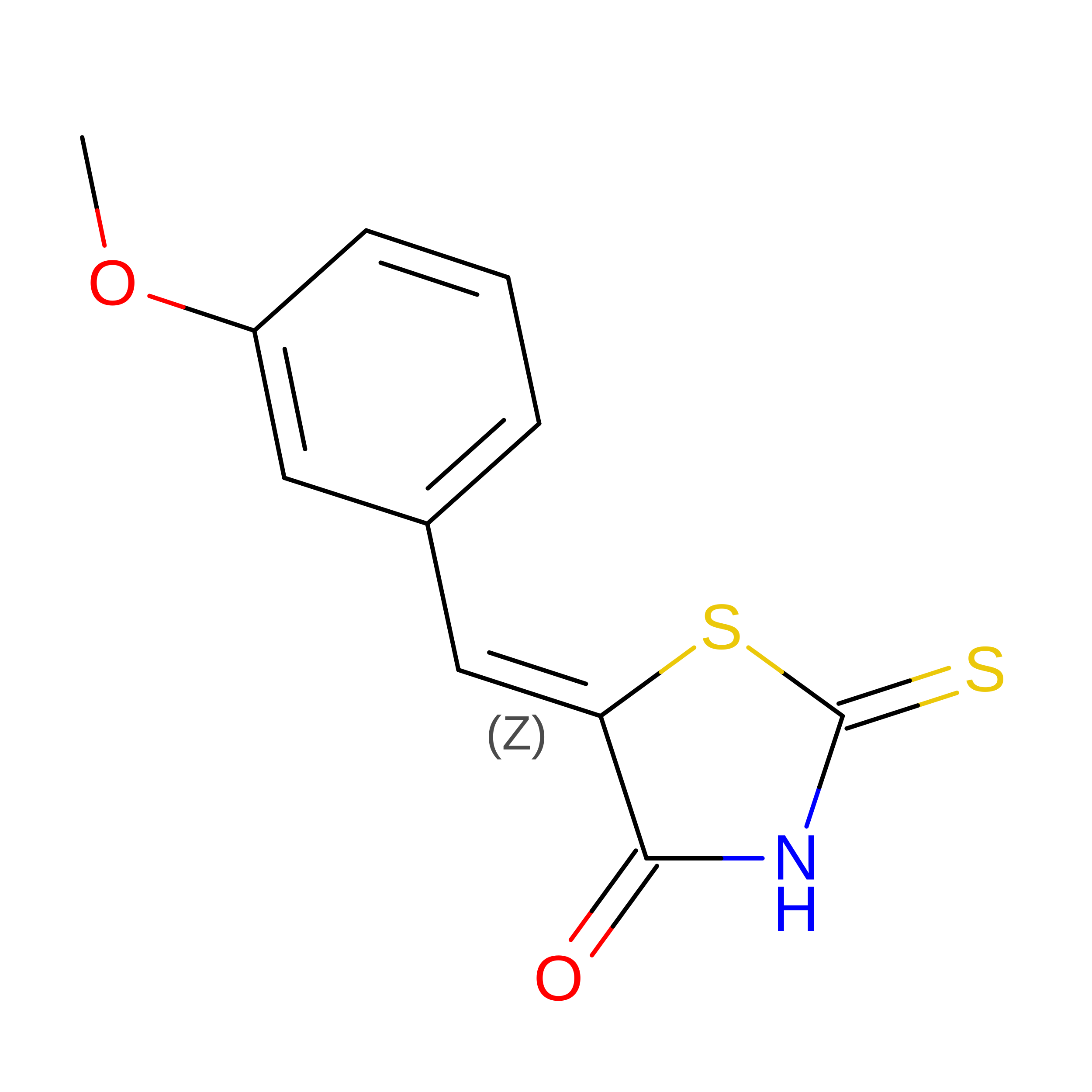
Mre11 dimer in complex with small molecule modulator PFMI


Experimental Data Snapshot

  • Method: X-RAY DIFFRACTION
  • Resolution: 2.35 Å
  • R-Value Free: 
    0.238 (Depositor), 0.240 (DCC) 
  • R-Value Work: 
    0.208 (Depositor), 0.210 (DCC) 
  • R-Value Observed: 
    0.210 (Depositor) 

Starting Model: experimental
View more details

wwPDB Validation 3D Report Full Report

Validation slider image for 6X1Y

Ligand Structure Quality Assessment 


This is version 1.3 of the entry. See complete history

Literature

Fragment- and structure-based drug discovery for developing therapeutic agents targeting the DNA Damage Response.

Wilson 3rd, D.M.Deacon, A.M.Duncton, M.A.J.Pellicena, P.Georgiadis, M.M.Yeh, A.P.Arvai, A.S.Moiani, D.Tainer, J.A.Das, D.

(2021) Prog Biophys Mol Biol 163: 130-142

  • DOI: https://doi.org/10.1016/j.pbiomolbio.2020.10.005
  • Primary Citation Related Structures: 
    6X1Y, 6X1Z

  • PubMed Abstract: 

    Cancer will directly affect the lives of over one-third of the population. The DNA Damage Response (DDR) is an intricate system involving damage recognition, cell cycle regulation, DNA repair, and ultimately cell fate determination, playing a central role in cancer etiology and therapy. Two primary therapeutic approaches involving DDR targeting include: combinatorial treatments employing anticancer genotoxic agents; and synthetic lethality, exploiting a sporadic DDR defect as a mechanism for cancer-specific therapy. Whereas, many DDR proteins have proven "undruggable", Fragment- and Structure-Based Drug Discovery (FBDD, SBDD) have advanced therapeutic agent identification and development. FBDD has led to 4 (with ∼50 more drugs under preclinical and clinical development), while SBDD is estimated to have contributed to the development of >200, FDA-approved medicines. Protein X-ray crystallography-based fragment library screening, especially for elusive or "undruggable" targets, allows for simultaneous generation of hits plus details of protein-ligand interactions and binding sites (orthosteric or allosteric) that inform chemical tractability, downstream biology, and intellectual property. Using a novel high-throughput crystallography-based fragment library screening platform, we screened five diverse proteins, yielding hit rates of ∼2-8% and crystal structures from ∼1.8 to 3.2 Å. We consider current FBDD/SBDD methods and some exemplary results of efforts to design inhibitors against the DDR nucleases meiotic recombination 11 (MRE11, a.k.a., MRE11A), apurinic/apyrimidinic endonuclease 1 (APE1, a.k.a., APEX1), and flap endonuclease 1 (FEN1).


  • Organizational Affiliation
    • Hasselt University, Biomedical Research Institute, Diepenbeek, Belgium; Boost Scientific, Heusden-Zolder, Belgium; XPose Therapeutics Inc., San Carlos, CA, USA.

Macromolecule Content 

  • Total Structure Weight: 78.04 kDa 
  • Atom Count: 5,389 
  • Modeled Residue Count: 654 
  • Deposited Residue Count: 672 
  • Unique protein chains: 1

Macromolecules

Find similar proteins by:|  3D Structure
Entity ID: 1
MoleculeChains  Sequence LengthOrganismDetailsImage
Nuclease SbcCD subunit D
A, B
336Thermotoga maritimaMutation(s): 0 
Gene Names: sbcDTM_1635
EC: 3.1
UniProt
Find proteins for Q9X1X0 (Thermotoga maritima (strain ATCC 43589 / DSM 3109 / JCM 10099 / NBRC 100826 / MSB8))
Explore Q9X1X0 
Go to UniProtKB:  Q9X1X0
Entity Groups
Sequence Clusters30% Identity50% Identity70% Identity90% Identity95% Identity100% Identity
UniProt GroupQ9X1X0
Sequence Annotations
Expand
Reference Sequence

Experimental Data & Validation

Experimental Data

  • Method: X-RAY DIFFRACTION
  • Resolution: 2.35 Å
  • R-Value Free:  0.238 (Depositor), 0.240 (DCC) 
  • R-Value Work:  0.208 (Depositor), 0.210 (DCC) 
  • R-Value Observed: 0.210 (Depositor) 
Space Group: P 1 21 1
Unit Cell:
Length ( Å )Angle ( ˚ )
a = 47.199α = 90
b = 109.147β = 99.934
c = 75.996γ = 90
Software Package:
Software NamePurpose
PHENIXrefinement
HKL-2000data reduction
HKL-2000data scaling
PHASERphasing

Structure Validation

View Full Validation Report


Ligand Structure Quality Assessment 


Entry History 

& Funding Information

Deposition Data


Funding OrganizationLocationGrant Number
National Institutes of Health/National Cancer Institute (NIH/NCI)United States5R01CA117638-15
National Institutes of Health/National Cancer Institute (NIH/NCI)United States5P01CA092584-19
National Institutes of Health/National Cancer Institute (NIH/NCI)United States5R35CA220430-02

Revision History  (Full details and data files)

  • Version 1.0: 2020-06-10
    Type: Initial release
  • Version 1.1: 2020-12-23
    Changes: Database references
  • Version 1.2: 2021-06-16
    Changes: Database references
  • Version 1.3: 2023-10-18
    Changes: Data collection, Database references, Refinement description