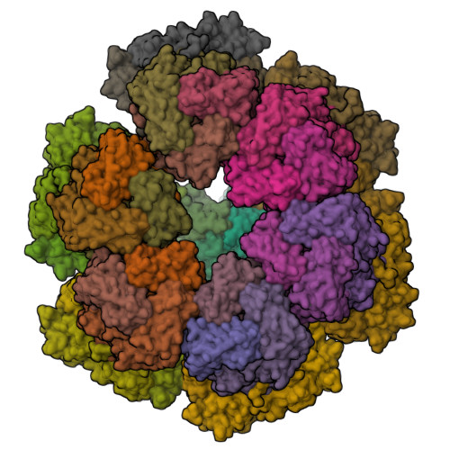
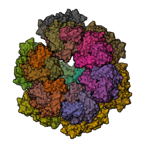
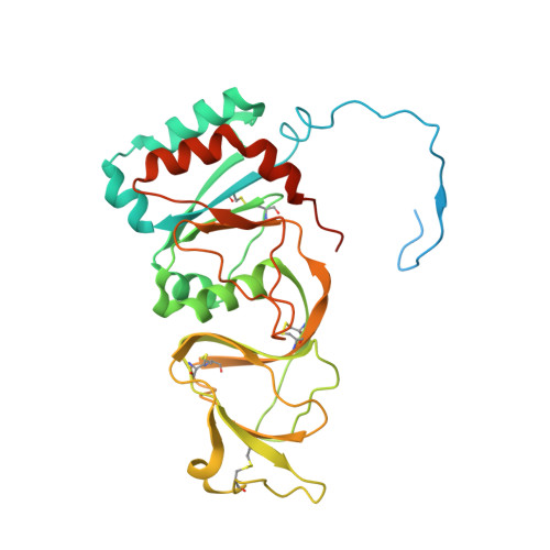
6WXG

Cryo-EM reconstruction of VP5*/VP8* assembly from rhesus rotavirus particles - Reversed conformation


Experimental Data Snapshot

  • Method: ELECTRON MICROSCOPY
  • Resolution: 3.30 Å
  • Aggregation State: PARTICLE 
  • Reconstruction Method: SINGLE PARTICLE 

Starting Models: experimental
View more details

wwPDB Validation   3D Report Full Report


This is version 1.3 of the entry. See complete history


Literature

Functional refolding of the penetration protein on a non-enveloped virus.

Herrmann, T.Torres, R.Salgado, E.N.Berciu, C.Stoddard, D.Nicastro, D.Jenni, S.Harrison, S.C.

(2021) Nature 590: 666-670

  • DOI: https://doi.org/10.1038/s41586-020-03124-4
  • Primary Citation of Related Structures:  
    6WXE, 6WXF, 6WXG

  • PubMed Abstract: 

    A non-enveloped virus requires a membrane lesion to deliver its genome into a target cell 1 . For rotaviruses, membrane perforation is a principal function of the viral outer-layer protein, VP4 2,3 . Here we describe theĀ use of electron cryomicroscopy to determine how VP4 performs this function and show that when activated by cleavage to VP8* and VP5*, VP4 can rearrange on the virion surface from an 'upright' to a 'reversed' conformation. The reversed structure projects a previously buried 'foot' domain outwards into the membrane of the host cell to which the virion has attached. Electron cryotomograms of virus particles entering cells are consistent with this picture. Using a disulfide mutant of VP4, we have also stabilized a probable intermediate in the transition between the two conformations. Our results define molecular mechanisms for the first steps of the penetration of rotaviruses into the membranes of target cells and suggest similarities with mechanisms postulated for other viruses.


  • Organizational Affiliation

    Department of Biological Chemistry and Molecular Pharmacology, Harvard Medical School, Boston, MA, USA.


Macromolecules
Find similar proteins by:  (by identity cutoff)  |  3D Structure
Entity ID: 1
MoleculeChains Sequence LengthOrganismDetailsImage
Outer capsid protein VP4A [auth 1],
B [auth 2],
C [auth 3]
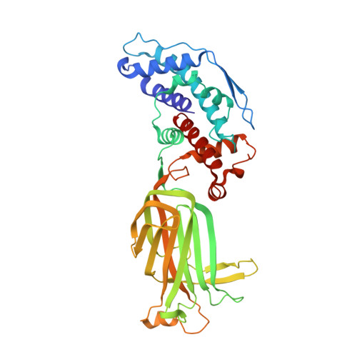
776Simian rotavirus A strain RRVMutation(s): 0 
UniProt
Find proteins for G0YZG6 (Rotavirus A (strain RVA/Monkey/United States/RRV/1975/G3P5B[3]))
Explore G0YZG6 
Go to UniProtKB:  G0YZG6
Entity Groups  
Sequence Clusters30% Identity50% Identity70% Identity90% Identity95% Identity100% Identity
UniProt GroupG0YZG6
Sequence Annotations
Expand
  • Reference Sequence
Find similar proteins by:  (by identity cutoff)  |  3D Structure
Entity ID: 2
MoleculeChains Sequence LengthOrganismDetailsImage
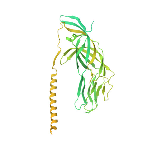
Intermediate capsid protein VP6397Simian rotavirus A strain RRVMutation(s): 0 
UniProt
Find proteins for B2BN53 (Rotavirus A (strain RVA/Monkey/United States/RRV/1975/G3P5B[3]))
Explore B2BN53 
Go to UniProtKB:  B2BN53
Entity Groups  
Sequence Clusters30% Identity50% Identity70% Identity90% Identity95% Identity100% Identity
UniProt GroupB2BN53
Sequence Annotations
Expand
  • Reference Sequence
Find similar proteins by:  (by identity cutoff)  |  3D Structure
Entity ID: 3
MoleculeChains Sequence LengthOrganismDetailsImage
Outer capsid glycoprotein VP7326Simian rotavirus A strain RRVMutation(s): 0 
UniProt
Find proteins for P12476 (Rotavirus A (strain RVA/Monkey/United States/RRV/1975/G3P5B[3]))
Explore P12476 
Go to UniProtKB:  P12476
Entity Groups  
Sequence Clusters30% Identity50% Identity70% Identity90% Identity95% Identity100% Identity
UniProt GroupP12476
Glycosylation
Glycosylation Sites: 1
Sequence Annotations
Expand
  • Reference Sequence
Small Molecules
Ligands 2 Unique
IDChains Name / Formula / InChI Key2D Diagram3D Interactions
NAG
Query on NAG

Download Ideal Coordinates CCD File 
BD [auth q]
DC [auth k]
FB [auth e]
FD [auth r]
HC [auth l]
BD [auth q],
DC [auth k],
FB [auth e],
FD [auth r],
HC [auth l],
JB [auth f],
JC [auth m],
LB [auth g],
NA [auth a],
PC [auth n],
RB [auth h],
TA [auth b],
TC [auth o],
VB [auth i],
VC [auth p],
XA [auth c],
XB [auth j],
ZA [auth d]
2-acetamido-2-deoxy-beta-D-glucopyranose
C8 H15 N O6
OVRNDRQMDRJTHS-FMDGEEDCSA-N
CA
Query on CA

Download Ideal Coordinates CCD File 
AB [auth d]
AC [auth j]
AD [auth p]
BB [auth d]
BC [auth j]
AB [auth d],
AC [auth j],
AD [auth p],
BB [auth d],
BC [auth j],
CB [auth d],
CC [auth j],
CD [auth q],
DB [auth d],
DD [auth q],
EB [auth d],
EC [auth k],
ED [auth q],
FC [auth k],
GB [auth e],
GC [auth k],
GD [auth r],
HB [auth e],
IB [auth e],
IC [auth l],
KB [auth f],
KC [auth m],
LC [auth m],
MB [auth g],
MC [auth m],
NB [auth g],
NC [auth m],
OA [auth a],
OB [auth g],
OC [auth m],
PA [auth a],
PB [auth g],
QA [auth a],
QB [auth g],
QC [auth n],
RA [auth a],
RC [auth n],
SA [auth a],
SB [auth h],
SC [auth n],
TB [auth h],
UA [auth b],
UB [auth h],
UC [auth o],
VA [auth b],
WA [auth b],
WB [auth i],
WC [auth p],
XC [auth p],
YA [auth c],
YB [auth j],
YC [auth p],
ZB [auth j],
ZC [auth p]
CALCIUM ION
Ca
BHPQYMZQTOCNFJ-UHFFFAOYSA-N
Experimental Data & Validation

Experimental Data

  • Method: ELECTRON MICROSCOPY
  • Resolution: 3.30 Å
  • Aggregation State: PARTICLE 
  • Reconstruction Method: SINGLE PARTICLE 
EM Software:
TaskSoftware PackageVersion
MODEL REFINEMENTPHENIX1.17.1-3660
RECONSTRUCTIONcisTEM

Structure Validation

View Full Validation Report


Entry History & Funding Information

Deposition Data


Funding OrganizationLocationGrant Number
National Institutes of Health/National Institute of General Medical Sciences (NIH/NIGMS)United StatesCA-13202

Revision History  (Full details and data files)

  • Version 1.0: 2021-01-20
    Type: Initial release
  • Version 1.1: 2021-01-27
    Changes: Database references
  • Version 1.2: 2021-03-10
    Changes: Database references
  • Version 1.3: 2024-11-13
    Changes: Data collection, Database references, Refinement description, Structure summary