6WX8 | pdb_00006wx8

SOX2 bound to Importin-alpha 3


Experimental Data Snapshot

  • Method: X-RAY DIFFRACTION
  • Resolution: 2.30 Å
  • R-Value Free: 
    0.245 (Depositor), 0.245 (DCC) 
  • R-Value Work: 
    0.220 (Depositor), 0.221 (DCC) 
  • R-Value Observed: 
    0.222 (Depositor) 

Starting Model: experimental
View more details

wwPDB Validation 3D Report Full Report

Validation slider image for 6WX8

This is version 1.3 of the entry. See complete history

Literature

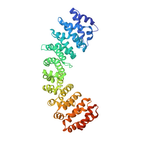
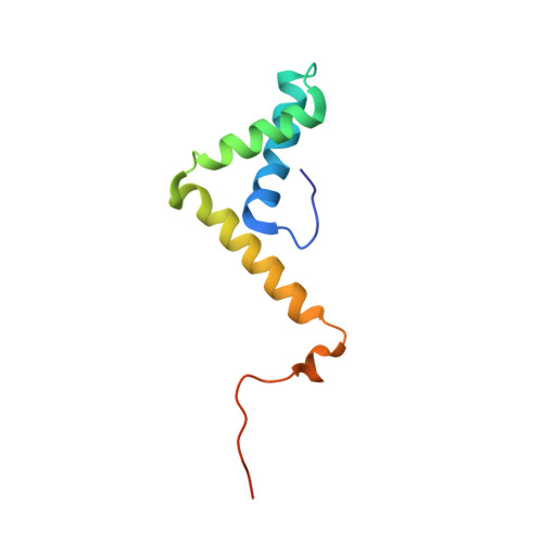
Structural basis for nuclear import selectivity of pioneer transcription factor SOX2.

Jagga, B.Edwards, M.Pagin, M.Wagstaff, K.M.Aragao, D.Roman, N.Nanson, J.D.Raidal, S.R.Dominado, N.Stewart, M.Jans, D.A.Hime, G.R.Nicolis, S.K.Basler, C.F.Forwood, J.K.

(2021) Nat Commun 12: 28-28

  • DOI: https://doi.org/10.1038/s41467-020-20194-0
  • Primary Citation Related Structures: 
    6WX7, 6WX8, 6WX9

  • PubMed Abstract: 

    SOX (SRY-related HMG-box) transcription factors perform critical functions in development and cell differentiation. These roles depend on precise nuclear trafficking, with mutations in the nuclear targeting regions causing developmental diseases and a range of cancers. SOX protein nuclear localization is proposed to be mediated by two nuclear localization signals (NLSs) positioned within the extremities of the DNA-binding HMG-box domain and, although mutations within either cause disease, the mechanistic basis has remained unclear. Unexpectedly, we find here that these two distantly positioned NLSs of SOX2 contribute to a contiguous interface spanning 9 of the 10 ARM domains on the nuclear import adapter IMPα3. We identify key binding determinants and show this interface is critical for neural stem cell maintenance and for Drosophila development. Moreover, we identify a structural basis for the preference of SOX2 binding to IMPα3. In addition to defining the structural basis for SOX protein localization, these results provide a platform for understanding how mutations and post-translational modifications within these regions may modulate nuclear localization and result in clinical disease, and also how other proteins containing multiple NLSs may bind IMPα through an extended recognition interface.


  • Organizational Affiliation
    • School of Biomedical Sciences, Charles Sturt University, Wagga Wagga, NSW, 2678, Australia.

Macromolecule Content 

  • Total Structure Weight: 123.22 kDa 
  • Atom Count: 8,113 
  • Modeled Residue Count: 999 
  • Deposited Residue Count: 1,098 
  • Unique protein chains: 2

Macromolecules

Find similar proteins by:|  3D Structure
Entity ID: 1
MoleculeChains  Sequence LengthOrganismDetailsImage
Importin subunit alpha-3
A, C
459Homo sapiensMutation(s): 0 
Gene Names: KPNA4QIP1
UniProt & NIH Common Fund Data Resources
Find proteins for O00629 (Homo sapiens)
Explore O00629 
Go to UniProtKB:  O00629
PHAROS:  O00629
GTEx:  ENSG00000186432 
Entity Groups
Sequence Clusters30% Identity50% Identity70% Identity90% Identity95% Identity100% Identity
UniProt GroupO00629
Sequence Annotations
Expand
Reference Sequence
Find similar proteins by:|  3D Structure
Entity ID: 2
MoleculeChains  Sequence LengthOrganismDetailsImage
Transcription factor SOX-2
B, D
90Homo sapiensMutation(s): 0 
Gene Names: SOX2
UniProt & NIH Common Fund Data Resources
Find proteins for P48431 (Homo sapiens)
Explore P48431 
Go to UniProtKB:  P48431
PHAROS:  P48431
GTEx:  ENSG00000181449 
Entity Groups
Sequence Clusters30% Identity50% Identity70% Identity90% Identity95% Identity100% Identity
UniProt GroupP48431
Sequence Annotations
Expand
Reference Sequence

Experimental Data & Validation

Experimental Data

  • Method: X-RAY DIFFRACTION
  • Resolution: 2.30 Å
  • R-Value Free:  0.245 (Depositor), 0.245 (DCC) 
  • R-Value Work:  0.220 (Depositor), 0.221 (DCC) 
  • R-Value Observed: 0.222 (Depositor) 
Space Group: C 1 2 1
Unit Cell:
Length ( Å )Angle ( ˚ )
a = 148.58α = 90
b = 118.99β = 128.77
c = 94.87γ = 90
Software Package:
Software NamePurpose
PHENIXrefinement
Aimlessdata reduction
Aimlessdata scaling
PHASERphasing

Structure Validation

View Full Validation Report


Entry History 

Deposition Data

Revision History  (Full details and data files)

  • Version 1.0: 2020-10-28
    Type: Initial release
  • Version 1.1: 2021-01-20
    Changes: Database references
  • Version 1.2: 2023-03-29
    Changes: Database references, Structure summary
  • Version 1.3: 2023-10-25
    Changes: Data collection, Refinement description