6WW0

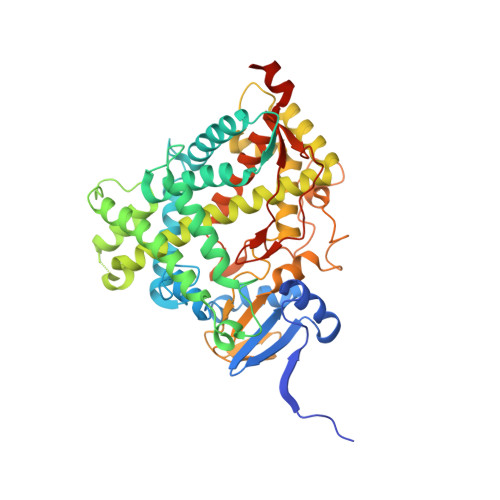
Human steroidogenic cytochrome P450 17A1 with 3-keto-5alpha-abiraterone analog


Experimental Data Snapshot

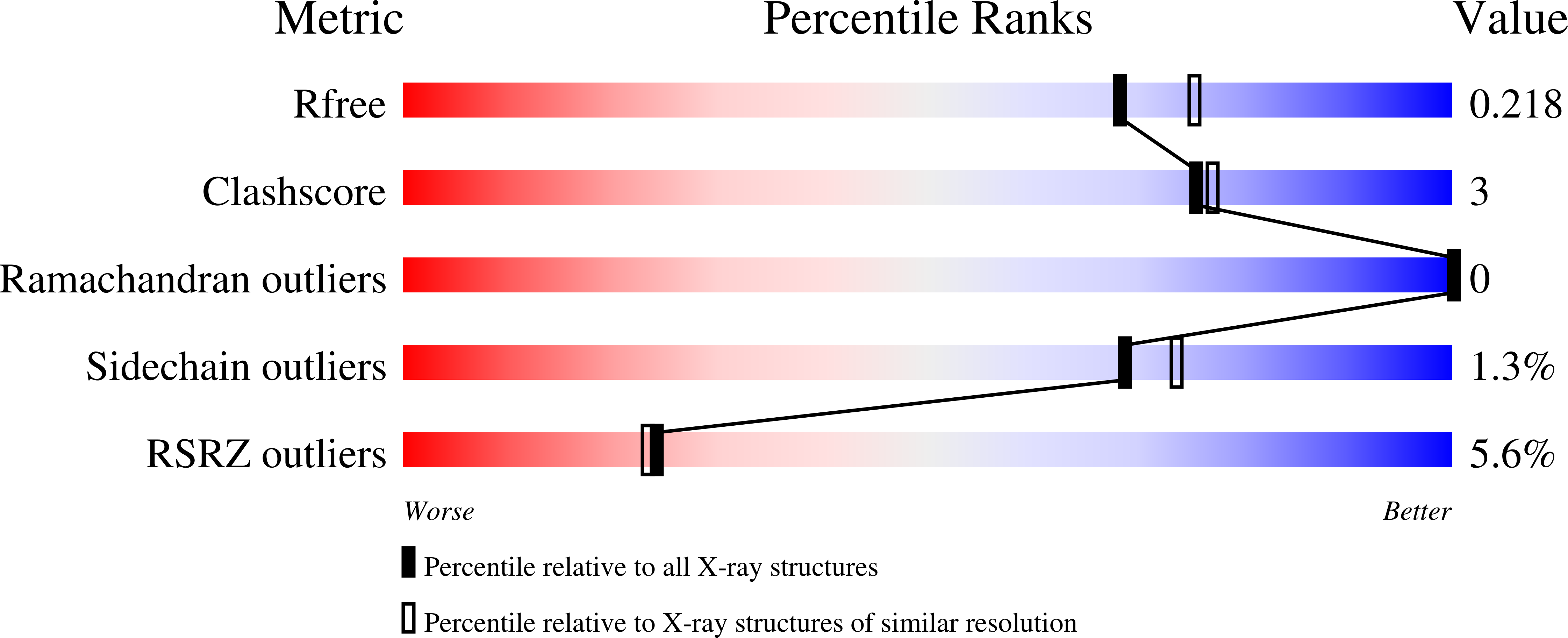
  • Method: X-RAY DIFFRACTION
  • Resolution: 2.01 Å
  • R-Value Free: 0.218 
  • R-Value Work: 0.185 
  • R-Value Observed: 0.187 

Starting Model: experimental
View more details

wwPDB Validation   3D Report Full Report


Ligand Structure Quality Assessment 


This is version 1.2 of the entry. See complete history


Literature

Human cytochrome P450 17A1 structures with metabolites of prostate cancer drug abiraterone reveal substrate-binding plasticity and a second binding site.

Petrunak, E.M.Bart, A.G.Peng, H.M.Auchus, R.J.Scott, E.E.

(2023) J Biol Chem 299: 102999-102999

  • DOI: https://doi.org/10.1016/j.jbc.2023.102999
  • Primary Citation of Related Structures:  
    5UYS, 6WR0, 6WR1, 6WW0

  • PubMed Abstract: 

    Abiraterone acetate is a first-line therapy for castration-resistant prostate cancer. This prodrug is deacetylated in vivo to abiraterone, which is a potent and specific inhibitor of cytochrome P450 17A1 (CYP17A1). CYP17A1 performs two sequential steps that are required for the biosynthesis of androgens that drive prostate cancer proliferation, analogous to estrogens in breast cancer. Abiraterone can be further metabolized in vivo on the steroid A ring to multiple metabolites that also inhibit CYP17A1. Despite its design as an active-site-directed substrate analog, abiraterone and its metabolites demonstrate mixed competitive/noncompetitive inhibition. To understand their binding, we solved the X-ray structures of CYP17A1 with three primary abiraterone metabolites. Despite different conformations of the steroid A ring and substituents, all three bound in the CYP17A1 active site with the steroid core packed against the I helix and the A ring C3 keto or hydroxyl oxygen forming a hydrogen bond with N202 similar to abiraterone itself. The structure of CYP17A1 with 3-keto, 5α-abiraterone was solved to 2.0 Å, the highest resolution to date for a CYP17A1 complex. This structure had additional electron density near the F/G loop, which is likely a second molecule of the inhibitor and which may explain the noncompetitive inhibition. Mutation of the adjacent Asn52 to Tyr positions its side chain in this space, maintains enzyme activity, and prevents binding of the peripheral ligand. Collectively, our findings provide further insight into abiraterone metabolite binding and CYP17A1 function.


  • Organizational Affiliation

    Department of Medicinal Chemistry, University of Kansas, Lawrence, Kansas, USA.


Macromolecules
Find similar proteins by:  (by identity cutoff)  |  3D Structure
Entity ID: 1
MoleculeChains Sequence LengthOrganismDetailsImage
Steroid 17-alpha-hydroxylase/17,20 lyase
A, B, C, D
494Homo sapiensMutation(s): 0 
Gene Names: CYP17A1CYP17S17AH
EC: 1.14.14.19 (PDB Primary Data), 1.14.14.32 (PDB Primary Data)
UniProt & NIH Common Fund Data Resources
Find proteins for P05093 (Homo sapiens)
Explore P05093 
Go to UniProtKB:  P05093
PHAROS:  P05093
GTEx:  ENSG00000148795 
Entity Groups  
Sequence Clusters30% Identity50% Identity70% Identity90% Identity95% Identity100% Identity
UniProt GroupP05093
Sequence Annotations
Expand
  • Reference Sequence
Small Molecules
Ligands 3 Unique
IDChains Name / Formula / InChI Key2D Diagram3D Interactions
HEM
Query on HEM

Download Ideal Coordinates CCD File 
E [auth A],
G [auth B],
I [auth C],
K [auth D]
PROTOPORPHYRIN IX CONTAINING FE
C34 H32 Fe N4 O4
KABFMIBPWCXCRK-RGGAHWMASA-L
UDJ (Subject of Investigation/LOI)
Query on UDJ

Download Ideal Coordinates CCD File 
F [auth A],
H [auth B],
J [auth C],
L [auth D]
(5alpha,8alpha)-17-(pyridin-3-yl)androst-16-en-3-one
C24 H31 N O
FKNZCFYWNHCQGE-IRMBCWQZSA-N
CL
Query on CL

Download Ideal Coordinates CCD File 
M [auth D],
N [auth D]
CHLORIDE ION
Cl
VEXZGXHMUGYJMC-UHFFFAOYSA-M
Experimental Data & Validation

Experimental Data

  • Method: X-RAY DIFFRACTION
  • Resolution: 2.01 Å
  • R-Value Free: 0.218 
  • R-Value Work: 0.185 
  • R-Value Observed: 0.187 
  • Space Group: P 21 21 21
Unit Cell:
Length ( Å )Angle ( ˚ )
a = 85.722α = 90
b = 151.182β = 90
c = 169.944γ = 90
Software Package:
Software NamePurpose
PHENIXrefinement
XDSdata reduction
SCALAdata scaling
PHASERphasing

Structure Validation

View Full Validation Report


Ligand Structure Quality Assessment 


Entry History & Funding Information

Deposition Data


Funding OrganizationLocationGrant Number
National Institutes of Health/National Institute of General Medical Sciences (NIH/NIGMS)United StatesR01 GM102505

Revision History  (Full details and data files)

  • Version 1.0: 2021-05-26
    Type: Initial release
  • Version 1.1: 2023-07-26
    Changes: Database references
  • Version 1.2: 2023-10-18
    Changes: Data collection, Refinement description