


Crystal structure of an aspartate ammonia-lyase from Elizabethkingia anophelis NUHP1
Edwards, T.E., Mayclin, S.J., Abendroth, J., Horanyi, P.S., Lorimer, D.D., Seattle Structural Genomics Center for Infectious Disease (SSGCID)To be published.
Experimental Data Snapshot
Starting Model: experimental
View more details
Entity ID: 1 | |||||
|---|---|---|---|---|---|
| Molecule | Chains | Sequence Length | Organism | Details | Image |
| Aspartate ammonia-lyase | 475 | Elizabethkingia anophelis NUH1 | Mutation(s): 0 Gene Names: BD94_1127 EC: 4.3.1.1 | 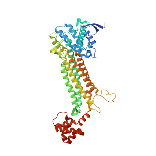 | |
UniProt | |||||
Find proteins for A0A077EBQ3 (Elizabethkingia anophelis NUHP1) Explore A0A077EBQ3 Go to UniProtKB: A0A077EBQ3 | |||||
Entity Groups | |||||
| Sequence Clusters | 30% Identity50% Identity70% Identity90% Identity95% Identity100% Identity | ||||
| UniProt Group | A0A077EBQ3 | ||||
Sequence AnnotationsExpand | |||||
Reference Sequence | |||||
| Ligands 4 Unique | |||||
|---|---|---|---|---|---|
| ID | Chains | Name / Formula / InChI Key | 2D Diagram | 3D Interactions | |
| TRS Download:Ideal Coordinates CCD File | E [auth A], K [auth B], Q [auth C], W [auth D] | 2-AMINO-2-HYDROXYMETHYL-PROPANE-1,3-DIOL C4 H12 N O3 LENZDBCJOHFCAS-UHFFFAOYSA-O |  | ||
| FUM (Subject of Investigation/LOI) Download:Ideal Coordinates CCD File | F [auth A], L [auth B], R [auth C], X [auth D] | FUMARIC ACID C4 H4 O4 VZCYOOQTPOCHFL-OWOJBTEDSA-N |  | ||
| GOL Download:Ideal Coordinates CCD File | G [auth A], M [auth B], S [auth C], Y [auth D] | GLYCEROL C3 H8 O3 PEDCQBHIVMGVHV-UHFFFAOYSA-N |  | ||
| CL Download:Ideal Coordinates CCD File | AA [auth D] BA [auth D] H [auth A] I [auth A] J [auth A] | CHLORIDE ION Cl VEXZGXHMUGYJMC-UHFFFAOYSA-M |  | ||
| Length ( Å ) | Angle ( ˚ ) |
|---|---|
| a = 92.82 | α = 90 |
| b = 96.85 | β = 90 |
| c = 212.2 | γ = 90 |
| Software Name | Purpose |
|---|---|
| XDS | data reduction |
| XSCALE | data scaling |
| MOLREP | phasing |
| PHENIX | refinement |
| PDB_EXTRACT | data extraction |


