6WIZ | pdb_00006wiz

Crystal structure of Fab 54-1G05 bound to H1 influenza hemagglutinin


Experimental Data Snapshot

  • Method: X-RAY DIFFRACTION
  • Resolution: 4.20 Å
  • R-Value Free: 
    0.389 (Depositor), 0.408 (DCC) 
  • R-Value Work: 
    0.323 (Depositor) 
  • R-Value Observed: 
    0.326 (Depositor) 

Starting Model: experimental
View more details

wwPDB Validation 3D Report Full Report

Validation slider image for 6WIZ

Ligand Structure Quality Assessment 


This is version 1.5 of the entry. See complete history

Literature

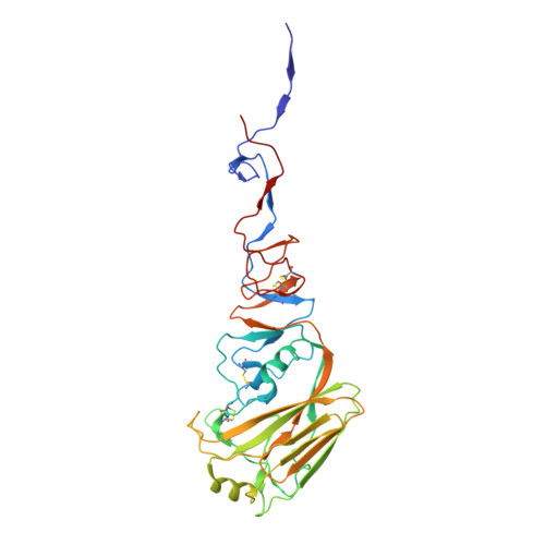
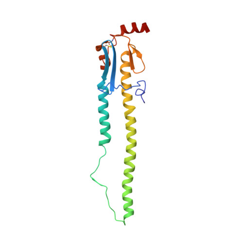
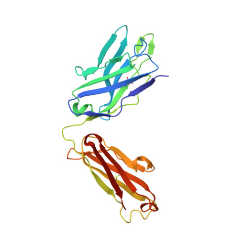
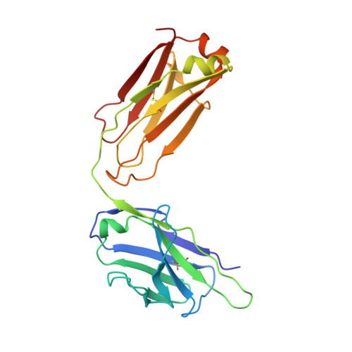
Convergent Evolution in Breadth of Two VH6-1-Encoded Influenza Antibody Clonotypes from a Single Donor.

Wu, N.C.Andrews, S.F.Raab, J.E.O'Connell, S.Schramm, C.A.Ding, X.Chambers, M.J.Leung, K.Wang, L.Zhang, Y.Mascola, J.R.Douek, D.C.Ledgerwood, J.E.McDermott, A.B.Wilson, I.A.

(2020) Cell Host Microbe 28: 434

  • DOI: https://doi.org/10.1016/j.chom.2020.06.003
  • Primary Citation Related Structures: 
    6WIY, 6WIZ, 6WJ0, 6WJ1

  • PubMed Abstract: 

    Understanding how broadly neutralizing antibodies (bnAbs) to influenza hemagglutinin (HA) naturally develop in humans is critical to the design of universal influenza vaccines. Several classes of bnAbs directed to the conserved HA stem were found in multiple individuals, including one encoded by heavy-chain variable domain V H 6-1. We describe two genetically similar V H 6-1 bnAb clonotypes from the same individual that exhibit different developmental paths toward broad neutralization activity. One clonotype evolved from a germline precursor recognizing influenza group 1 subtypes to gain breadth to group 2 subtypes. The other clonotype recognized group 2 subtypes and developed binding to group 1 subtypes through somatic hypermutation. Crystal structures reveal that the specificity differences are primarily mediated by complementarity-determining region H3 (CDR H3). Thus, while V H 6-1 provides a framework for development of HA stem-directed bnAbs, sequence differences in CDR H3 junctional regions during VDJ recombination can alter reactivity and evolutionary pathways toward increased breadth.


  • Organizational Affiliation
    • Department of Integrative Structural and Computational Biology, The Scripps Research Institute, San Diego, CA 92037, USA.

Macromolecule Content 

  • Total Structure Weight: 104.97 kDa 
  • Atom Count: 7,442 
  • Modeled Residue Count: 938 
  • Deposited Residue Count: 948 
  • Unique protein chains: 4

Macromolecules

Find similar proteins by:|  3D Structure
Entity ID: 1
MoleculeChains  Sequence LengthOrganismDetailsImage
Hemagglutinin HA1327Influenza A virusMutation(s): 0 
UniProt
Find proteins for A7Y8I1 (Influenza A virus)
Explore A7Y8I1 
Go to UniProtKB:  A7Y8I1
Entity Groups
Sequence Clusters30% Identity50% Identity70% Identity90% Identity95% Identity100% Identity
UniProt GroupA7Y8I1
Glycosylation
Glycosylation Sites: 1
Sequence Annotations
Expand
Reference Sequence
Find similar proteins by:|  3D Structure
Entity ID: 2
MoleculeChains  Sequence LengthOrganismDetailsImage
Hemagglutinin HA2174Influenza A virusMutation(s): 0 
UniProt
Find proteins for A7UPX0 (Influenza A virus)
Explore A7UPX0 
Go to UniProtKB:  A7UPX0
Entity Groups
Sequence Clusters30% Identity50% Identity70% Identity90% Identity95% Identity100% Identity
UniProt GroupA7UPX0
Sequence Annotations
Expand
Reference Sequence
Find similar proteins by:|  3D Structure
Entity ID: 3
MoleculeChains  Sequence LengthOrganismDetailsImage
Fab 54-1G05 heavy chainC [auth H]230Homo sapiensMutation(s): 0 
Find similar proteins by:|  3D Structure
Entity ID: 4
MoleculeChains  Sequence LengthOrganismDetailsImage
Fab 54-1G05 light chainD [auth L]217Homo sapiensMutation(s): 0 

Small Molecules

Ligands 1 Unique
IDChains Name / Formula / InChI Key2D Diagram3D Interactions
NAG

Query on NAG


Download:Ideal Coordinates CCD File
E [auth A]2-acetamido-2-deoxy-beta-D-glucopyranose
C8 H15 N O6
OVRNDRQMDRJTHS-FMDGEEDCSA-N

Experimental Data & Validation

Experimental Data

  • Method: X-RAY DIFFRACTION
  • Resolution: 4.20 Å
  • R-Value Free:  0.389 (Depositor), 0.408 (DCC) 
  • R-Value Work:  0.323 (Depositor) 
  • R-Value Observed: 0.326 (Depositor) 
Space Group: P 3 2 1
Unit Cell:
Length ( Å )Angle ( ˚ )
a = 187.378α = 90
b = 187.378β = 90
c = 132.533γ = 120
Software Package:
Software NamePurpose
PHENIXrefinement
Aimlessdata scaling
PDB_EXTRACTdata extraction
PHASERphasing

Structure Validation

View Full Validation Report


Ligand Structure Quality Assessment 


Entry History 

& Funding Information

Deposition Data


Funding OrganizationLocationGrant Number
National Institutes of Health/National Institute Of Allergy and Infectious Diseases (NIH/NIAID)United StatesR56 AI127371
National Institutes of Health/National Institute Of Allergy and Infectious Diseases (NIH/NIAID)United StatesK99 AI139445

Revision History  (Full details and data files)

  • Version 1.0: 2020-07-01
    Type: Initial release
  • Version 1.1: 2020-07-15
    Changes: Database references
  • Version 1.2: 2020-07-29
    Type: Remediation
    Reason: Carbohydrate remediation
    Changes: Data collection, Derived calculations, Structure summary
  • Version 1.3: 2020-09-23
    Changes: Database references, Structure summary
  • Version 1.4: 2023-10-18
    Changes: Advisory, Data collection, Database references, Refinement description
  • Version 1.5: 2024-11-06
    Changes: Structure summary