6W9L

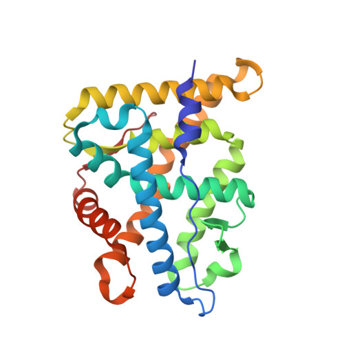
Structure of the Ancestral Glucocorticoid Receptor 2 ligand binding domain in complex with deacetylated deflazacort and PGC1a coregulator fragment

  • Classification: HORMONE
  • Organism(s): synthetic construct, Homo sapiens
  • Expression System: Escherichia coli
  • Mutation(s): No 

  • Deposited: 2020-03-23 Released: 2020-11-04 
  • Deposition Author(s): Liu, X., Ortlund, E.A.
  • Funding Organization(s): American Heart Association, National Institutes of Health/National Institute of Diabetes and Digestive and Kidney Disease (NIH/NIDDK)

Experimental Data Snapshot

  • Method: X-RAY DIFFRACTION
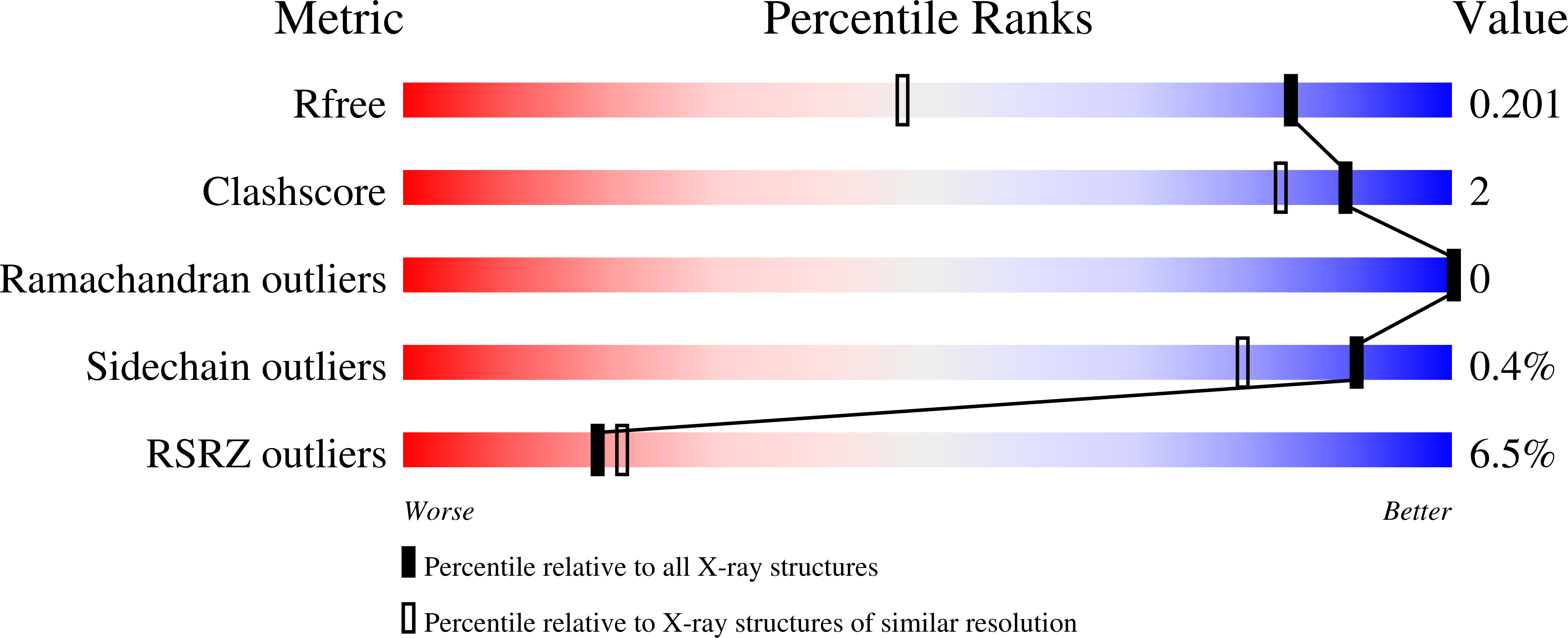
  • Resolution: 1.45 Å
  • R-Value Free: 0.200 
  • R-Value Work: 0.174 
  • R-Value Observed: 0.175 

Starting Model: experimental
View more details

wwPDB Validation   3D Report Full Report


Ligand Structure Quality Assessment 


This is version 1.1 of the entry. See complete history


Literature

Disruption of a key ligand-H-bond network drives dissociative properties in vamorolone for Duchenne muscular dystrophy treatment.

Liu, X.Wang, Y.Gutierrez, J.S.Damsker, J.M.Nagaraju, K.Hoffman, E.P.Ortlund, E.A.

(2020) Proc Natl Acad Sci U S A 117: 24285-24293

  • DOI: https://doi.org/10.1073/pnas.2006890117
  • Primary Citation of Related Structures:  
    6W9K, 6W9L, 6W9M

  • PubMed Abstract: 

    Duchenne muscular dystrophy is a genetic disorder that shows chronic and progressive damage to skeletal and cardiac muscle leading to premature death. Antiinflammatory corticosteroids targeting the glucocorticoid receptor (GR) are the current standard of care but drive adverse side effects such as deleterious bone loss. Through subtle modification to a steroidal backbone, a recently developed drug, vamorolone, appears to preserve beneficial efficacy but with significantly reduced side effects. We use combined structural, biophysical, and biochemical approaches to show that loss of a receptor-ligand hydrogen bond drives these remarkable therapeutic effects. Moreover, vamorolone uniformly weakens coactivator associations but not corepressor associations, implicating partial agonism as the main driver of its dissociative properties. Additionally, we identify a critical and evolutionarily conserved intramolecular network connecting the ligand to the coregulator binding surface. Interruption of this allosteric network by vamorolone selectively reduces GR-driven transactivation while leaving transrepression intact. Our results establish a mechanistic understanding of how vamorolone reduces side effects, guiding the future design of partial agonists as selective GR modulators with an improved therapeutic index.


  • Organizational Affiliation

    Department of Biochemistry, Emory University, Atlanta, GA 30322.


Macromolecules
Find similar proteins by:  (by identity cutoff)  |  3D Structure
Entity ID: 1
MoleculeChains Sequence LengthOrganismDetailsImage
Glucocorticoid Receptor249synthetic constructMutation(s): 0 
UniProt
Find proteins for A0A1X8XLE9 (unidentified)
Explore A0A1X8XLE9 
Go to UniProtKB:  A0A1X8XLE9
Entity Groups  
Sequence Clusters30% Identity50% Identity70% Identity90% Identity95% Identity100% Identity
UniProt GroupA0A1X8XLE9
Sequence Annotations
Expand
  • Reference Sequence

Find similar proteins by:  Sequence   |   3D Structure  

Entity ID: 2
MoleculeChains Sequence LengthOrganismDetailsImage
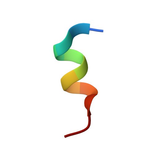
Peroxisome proliferator-activated receptor gamma coactivator 1-alpha12Homo sapiensMutation(s): 0 
UniProt & NIH Common Fund Data Resources
Find proteins for Q9UBK2 (Homo sapiens)
Explore Q9UBK2 
Go to UniProtKB:  Q9UBK2
PHAROS:  Q9UBK2
GTEx:  ENSG00000109819 
Entity Groups  
Sequence Clusters30% Identity50% Identity70% Identity90% Identity95% Identity100% Identity
UniProt GroupQ9UBK2
Sequence Annotations
Expand
  • Reference Sequence
Small Molecules
Ligands 2 Unique
IDChains Name / Formula / InChI Key2D Diagram3D Interactions
TUS (Subject of Investigation/LOI)
Query on TUS

Download Ideal Coordinates CCD File 
C [auth A](4aR,4bS,5S,6aS,6bS,9aR,10aS,10bS)-5-hydroxy-6b-(hydroxyacetyl)-4a,6a,8-trimethyl-4a,4b,5,6,6a,6b,9a,10,10a,10b,11,12-dodecahydro-2H-naphtho[2',1':4,5]indeno[1,2-d][1,3]oxazol-2-one
C23 H29 N O5
KENSGCYKTRNIST-RVUAFKSESA-N
GOL
Query on GOL

Download Ideal Coordinates CCD File 
D [auth A]GLYCEROL
C3 H8 O3
PEDCQBHIVMGVHV-UHFFFAOYSA-N
Experimental Data & Validation

Experimental Data

  • Method: X-RAY DIFFRACTION
  • Resolution: 1.45 Å
  • R-Value Free: 0.200 
  • R-Value Work: 0.174 
  • R-Value Observed: 0.175 
  • Space Group: C 2 2 21
Unit Cell:
Length ( Å )Angle ( ˚ )
a = 71.691α = 90
b = 96.536β = 90
c = 107.46γ = 90
Software Package:
Software NamePurpose
PHENIXrefinement
HKL-2000data reduction
HKL-2000data scaling
PHASERphasing

Structure Validation

View Full Validation Report


Ligand Structure Quality Assessment 


Entry History & Funding Information

Deposition Data


Funding OrganizationLocationGrant Number
American Heart AssociationUnited States17POST33660110
National Institutes of Health/National Institute of Diabetes and Digestive and Kidney Disease (NIH/NIDDK)United StatesR01DK095750

Revision History  (Full details and data files)

  • Version 1.0: 2020-11-04
    Type: Initial release
  • Version 1.1: 2023-10-18
    Changes: Data collection, Database references, Refinement description