6W90 | pdb_00006w90

De novo designed NTF2 fold protein NT-9


Experimental Data Snapshot

  • Method: X-RAY DIFFRACTION
  • Resolution: 1.50 Å
  • R-Value Free: 
    0.210 (Depositor), 0.228 (DCC) 
  • R-Value Work: 
    0.186 (Depositor) 
  • R-Value Observed: 
    0.188 (Depositor) 

Starting Model: in silico
View more details

wwPDB Validation 3D Report Full Report

Validation slider image for 6W90

Ligand Structure Quality Assessment 


This is version 2.1 of the entry. See complete history

Literature

Expanding the space of protein geometries by computational design of de novo fold families.

Pan, X.Thompson, M.C.Zhang, Y.Liu, L.Fraser, J.S.Kelly, M.J.S.Kortemme, T.

(2020) Science 369: 1132-1136

  • DOI: https://doi.org/10.1126/science.abc0881
  • Primary Citation Related Structures: 
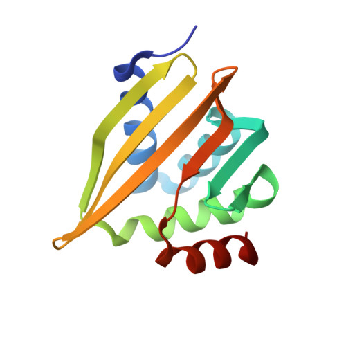
    6VG7, 6VGA, 6VGB, 6W90

  • PubMed Abstract: 

    Naturally occurring proteins vary the precise geometries of structural elements to create distinct shapes optimal for function. We present a computational design method, loop-helix-loop unit combinatorial sampling (LUCS), that mimics nature's ability to create families of proteins with the same overall fold but precisely tunable geometries. Through near-exhaustive sampling of loop-helix-loop elements, LUCS generates highly diverse geometries encompassing those found in nature but also surpassing known structure space. Biophysical characterization showed that 17 (38%) of 45 tested LUCS designs encompassing two different structural topologies were well folded, including 16 with designed non-native geometries. Four experimentally solved structures closely matched the designs. LUCS greatly expands the designable structure space and offers a new paradigm for designing proteins with tunable geometries that may be customizable for novel functions.


  • Organizational Affiliation
    • Department of Bioengineering and Therapeutic Sciences, University of California, San Francisco, CA, USA. xingjiepan@gmail.com tanjakortemme@gmail.com.

Macromolecule Content 

  • Total Structure Weight: 16.53 kDa 
  • Atom Count: 1,344 
  • Modeled Residue Count: 130 
  • Deposited Residue Count: 131 
  • Unique protein chains: 1

Macromolecules

Find similar proteins by:|  3D Structure
Entity ID: 1
MoleculeChains  Sequence LengthOrganismDetailsImage
NTF2 fold protein loop-helix-loop design NT-9131synthetic constructMutation(s): 0 

Small Molecules

Ligands 1 Unique
IDChains Name / Formula / InChI Key2D Diagram3D Interactions
3PE

Query on 3PE


Download:Ideal Coordinates CCD File
B [auth A]1,2-Distearoyl-sn-glycerophosphoethanolamine
C41 H82 N O8 P
LVNGJLRDBYCPGB-LDLOPFEMSA-N

Experimental Data & Validation

Experimental Data

  • Method: X-RAY DIFFRACTION
  • Resolution: 1.50 Å
  • R-Value Free:  0.210 (Depositor), 0.228 (DCC) 
  • R-Value Work:  0.186 (Depositor) 
  • R-Value Observed: 0.188 (Depositor) 
Space Group: P 21 21 21
Unit Cell:
Length ( Å )Angle ( ˚ )
a = 33.5α = 90
b = 51.99β = 90
c = 66.41γ = 90
Software Package:
Software NamePurpose
PHENIXrefinement
xia2data reduction
xia2data scaling
PHASERphasing

Structure Validation

View Full Validation Report


Ligand Structure Quality Assessment 


Entry History 

& Funding Information

Deposition Data


Funding OrganizationLocationGrant Number
National Institutes of Health/National Institute of General Medical Sciences (NIH/NIGMS)United StatesR01GM110089

Revision History  (Full details and data files)

  • Version 1.0: 2020-08-19
    Type: Initial release
  • Version 1.1: 2020-09-09
    Changes: Database references
  • Version 2.0: 2024-03-06
    Changes: Atomic model, Data collection, Database references, Derived calculations, Structure summary
  • Version 2.1: 2024-04-03
    Changes: Refinement description