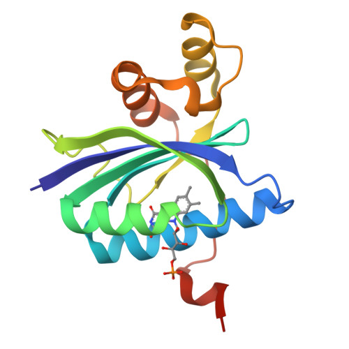
6W6Z

BlsA ground state

  • Classification: FLAVOPROTEIN
  • Organism(s): Acinetobacter baumannii
  • Expression System: Escherichia coli
  • Mutation(s): No 

  • Deposited: 2020-03-18 Released: 2020-10-07 
  • Deposition Author(s): Chitrakar, I., French, J.B.
  • Funding Organization(s): National Institutes of Health/National Institute of General Medical Sciences (NIH/NIGMS), National Science Foundation (NSF, United States)

Experimental Data Snapshot

  • Method: X-RAY DIFFRACTION
  • Resolution: 1.71 Å
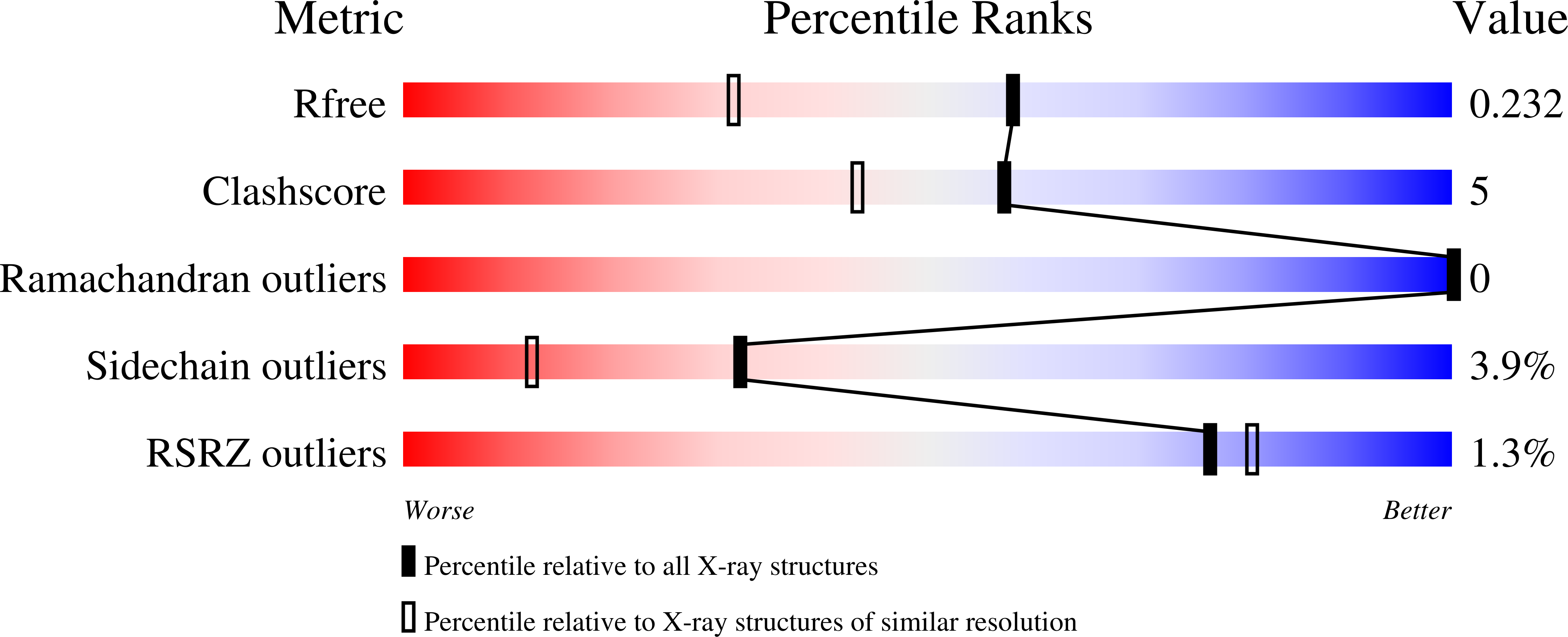
  • R-Value Free: 0.222 
  • R-Value Work: 0.185 
  • R-Value Observed: 0.187 

Starting Model: experimental
View more details

wwPDB Validation   3D Report Full Report


Ligand Structure Quality Assessment 


This is version 1.2 of the entry. See complete history


Literature

Structural Basis for the Regulation of Biofilm Formation and Iron Uptake in A. baumannii by the Blue-Light-Using Photoreceptor, BlsA.

Chitrakar, I.Iuliano, J.N.He, Y.Woroniecka, H.A.Tolentino Collado, J.Wint, J.M.Walker, S.G.Tonge, P.J.French, J.B.

(2020) ACS Infect Dis 6: 2592-2603

  • DOI: https://doi.org/10.1021/acsinfecdis.0c00156
  • Primary Citation of Related Structures:  
    6W6Z, 6W72

  • PubMed Abstract: 

    The opportunistic human pathogen, A. baumannii , senses and responds to light using the blue light sensing A (BlsA) photoreceptor protein. BlsA is a blue-light-using flavin adenine dinucleotide (BLUF) protein that is known to regulate a wide variety of cellular functions through interactions with different binding partners. Using immunoprecipitation of tagged BlsA in A. baumannii lysates, we observed a number of proteins that interact with BlsA, including several transcription factors. In addition to a known binding partner, the iron uptake regulator Fur, we identified the biofilm response regulator BfmR as a putative BlsA-binding partner. Using microscale thermophoresis, we determined that both BfmR and Fur bind to BlsA with nanomolar binding constants. To better understand how BlsA interacts with and regulates these transcription factors, we solved the X-ray crystal structures of BlsA in both a ground (dark) state and a photoactivated light state. Comparison of the light- and dark-state structures revealed that, upon photoactivation, the two α-helices comprising the variable domain of BlsA undergo a distinct conformational change. The flavin-binding site, however, remains largely unchanged from dark to light. These structures, along with docking studies of BlsA and Fur, reveal key mechanistic details about how BlsA propagates the photoactivation signal between protein domains and on to its binding partner. Taken together, our structural and biophysical data provide important insights into how BlsA controls signal transduction in A. baumannii and provides a likely mechanism for blue-light-dependent modulation of biofilm formation and iron uptake.


  • Organizational Affiliation

    The Hormel Institute, University of Minnesota, Austin, Minnesota 55912, United States.


Macromolecules
Find similar proteins by:  (by identity cutoff)  |  3D Structure
Entity ID: 1
MoleculeChains Sequence LengthOrganismDetailsImage
BLUF domain-containing protein156Acinetobacter baumanniiMutation(s): 0 
Gene Names: 
UniProt
Find proteins for V5VB82 (Acinetobacter baumannii)
Explore V5VB82 
Go to UniProtKB:  V5VB82
Entity Groups  
Sequence Clusters30% Identity50% Identity70% Identity90% Identity95% Identity100% Identity
UniProt GroupV5VB82
Sequence Annotations
Expand
  • Reference Sequence
Small Molecules
Ligands 1 Unique
IDChains Name / Formula / InChI Key2D Diagram3D Interactions
FMN
Query on FMN

Download Ideal Coordinates CCD File 
B [auth A]FLAVIN MONONUCLEOTIDE
C17 H21 N4 O9 P
FVTCRASFADXXNN-SCRDCRAPSA-N
Experimental Data & Validation

Experimental Data

  • Method: X-RAY DIFFRACTION
  • Resolution: 1.71 Å
  • R-Value Free: 0.222 
  • R-Value Work: 0.185 
  • R-Value Observed: 0.187 
  • Space Group: C 1 2 1
Unit Cell:
Length ( Å )Angle ( ˚ )
a = 92.873α = 90
b = 37.438β = 106.57
c = 49.269γ = 90
Software Package:
Software NamePurpose
REFMACrefinement
XDSdata reduction
Aimlessdata scaling
PDB_EXTRACTdata extraction
MOLREPphasing

Structure Validation

View Full Validation Report


Ligand Structure Quality Assessment 


Entry History & Funding Information

Deposition Data


Funding OrganizationLocationGrant Number
National Institutes of Health/National Institute of General Medical Sciences (NIH/NIGMS)United StatesR35GM124898
National Institutes of Health/National Institute of General Medical Sciences (NIH/NIGMS)United StatesT32GM092714
National Institutes of Health/National Institute of General Medical Sciences (NIH/NIGMS)United StatesR25GM103962
National Science Foundation (NSF, United States)United StatesMCB1750637
National Science Foundation (NSF, United States)United StatesCHE1223819

Revision History  (Full details and data files)

  • Version 1.0: 2020-10-07
    Type: Initial release
  • Version 1.1: 2020-10-21
    Changes: Database references
  • Version 1.2: 2023-10-18
    Changes: Data collection, Database references, Refinement description