6VWW | pdb_00006vww

Crystal Structure of NSP15 Endoribonuclease from SARS CoV-2.


Experimental Data Snapshot

  • Method: X-RAY DIFFRACTION
  • Resolution: 2.20 Å
  • R-Value Free: 
    0.178 (Depositor), 0.175 (DCC) 
  • R-Value Work: 
    0.158 (Depositor), 0.156 (DCC) 
  • R-Value Observed: 
    0.161 (Depositor) 

Starting Model: experimental
View more details

wwPDB Validation 3D Report Full Report

Validation slider image for 6VWW

This is version 2.2 of the entry. See complete history

Literature

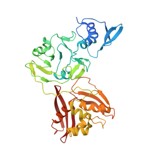
Crystal structure of Nsp15 endoribonuclease NendoU from SARS-CoV-2.

Kim, Y.Jedrzejczak, R.Maltseva, N.I.Wilamowski, M.Endres, M.Godzik, A.Michalska, K.Joachimiak, A.

(2020) Protein Sci 29: 1596-1605

  • DOI: https://doi.org/10.1002/pro.3873
  • Primary Citation Related Structures: 
    6VWW, 6W01

  • PubMed Abstract: 

    Severe Acute Respiratory Syndrome coronavirus 2 (SARS-CoV-2) is rapidly spreading around the world. There is no existing vaccine or proven drug to prevent infections and stop virus proliferation. Although this virus is similar to human and animal SARS-CoVs and Middle East Respiratory Syndrome coronavirus (MERS-CoVs), the detailed information about SARS-CoV-2 proteins structures and functions is urgently needed to rapidly develop effective vaccines, antibodies, and antivirals. We applied high-throughput protein production and structure determination pipeline at the Center for Structural Genomics of Infectious Diseases to produce SARS-CoV-2 proteins and structures. Here we report two high-resolution crystal structures of endoribonuclease Nsp15/NendoU. We compare these structures with previously reported homologs from SARS and MERS coronaviruses.


  • Organizational Affiliation
    • Center for Structural Genomics of Infectious Diseases, Consortium for Advanced Science and Engineering, University of Chicago, Chicago, Illinois, USA.

Macromolecule Content 

  • Total Structure Weight: 84.38 kDa 
  • Atom Count: 6,009 
  • Modeled Residue Count: 696 
  • Deposited Residue Count: 740 
  • Unique protein chains: 1

Macromolecules

Find similar proteins by:|  3D Structure
Entity ID: 1
MoleculeChains  Sequence LengthOrganismDetailsImage
Uridylate-specific endoribonuclease
A, B
370Severe acute respiratory syndrome coronavirus 2Mutation(s): 0 
Gene Names: rep1a-1b
EC: 3.1
UniProt
Find proteins for P0DTD1 (Severe acute respiratory syndrome coronavirus 2)
Explore P0DTD1 
Go to UniProtKB:  P0DTD1
Entity Groups
Sequence Clusters30% Identity50% Identity70% Identity90% Identity95% Identity100% Identity
UniProt GroupP0DTD1
Sequence Annotations
Expand
Reference Sequence

Small Molecules

Ligands 4 Unique
IDChains Name / Formula / InChI Key2D Diagram3D Interactions
GOL

Query on GOL


Download:Ideal Coordinates CCD File
C [auth A]
D [auth A]
F [auth A]
G [auth A]
I [auth B]
C [auth A],
D [auth A],
F [auth A],
G [auth A],
I [auth B],
J [auth B],
K [auth B],
M [auth B],
N [auth B]
GLYCEROL
C3 H8 O3
PEDCQBHIVMGVHV-UHFFFAOYSA-N
ACY

Query on ACY


Download:Ideal Coordinates CCD File
H [auth A],
O [auth B],
P [auth B]
ACETIC ACID
C2 H4 O2
QTBSBXVTEAMEQO-UHFFFAOYSA-N
CL

Query on CL


Download:Ideal Coordinates CCD File
L [auth B]CHLORIDE ION
Cl
VEXZGXHMUGYJMC-UHFFFAOYSA-M
MG

Query on MG


Download:Ideal Coordinates CCD File
E [auth A]MAGNESIUM ION
Mg
JLVVSXFLKOJNIY-UHFFFAOYSA-N

Experimental Data & Validation

Experimental Data

  • Method: X-RAY DIFFRACTION
  • Resolution: 2.20 Å
  • R-Value Free:  0.178 (Depositor), 0.175 (DCC) 
  • R-Value Work:  0.158 (Depositor), 0.156 (DCC) 
  • R-Value Observed: 0.161 (Depositor) 
Space Group: P 63
Unit Cell:
Length ( Å )Angle ( ˚ )
a = 150.539α = 90
b = 150.539β = 90
c = 111.31γ = 120
Software Package:
Software NamePurpose
PHENIXrefinement
HKL-3000data reduction
HKL-3000data scaling
HKL-3000phasing
MOLREPphasing

Structure Validation

View Full Validation Report


Entry History 

& Funding Information

Deposition Data


Funding OrganizationLocationGrant Number
National Institutes of Health/National Institute Of Allergy and Infectious Diseases (NIH/NIAID)United States--

Revision History  (Full details and data files)

  • Version 1.0: 2020-03-04
    Type: Initial release
  • Version 1.1: 2020-03-11
    Changes: Database references, Source and taxonomy
  • Version 1.2: 2020-04-01
    Changes: Data collection
  • Version 1.3: 2020-05-06
    Changes: Database references, Source and taxonomy, Structure summary
  • Version 2.0: 2020-06-10
    Changes: Advisory, Atomic model, Database references, Derived calculations, Polymer sequence, Source and taxonomy, Structure summary
  • Version 2.1: 2020-07-08
    Changes: Database references
  • Version 2.2: 2023-10-11
    Changes: Data collection, Database references, Refinement description