6VSU | pdb_00006vsu

Arginase from Arabidopsis thaliana in Complex with Ornithine


Experimental Data Snapshot

  • Method: X-RAY DIFFRACTION
  • Resolution: 2.25 Å
  • R-Value Free: 
    0.197 (Depositor), 0.202 (DCC) 
  • R-Value Work: 
    0.159 (Depositor), 0.167 (DCC) 
  • R-Value Observed: 
    0.159 (Depositor) 

Starting Model: experimental
View more details

wwPDB Validation 3D Report Full Report

Validation slider image for 6VSU

Ligand Structure Quality Assessment 


This is version 1.2 of the entry. See complete history

Literature

The Neighboring Subunit Is Engaged to Stabilize the Substrate in the Active Site of Plant Arginases.

Sekula, B.

(2020) Front Plant Sci 11: 987-987

  • DOI: https://doi.org/10.3389/fpls.2020.00987
  • Primary Citation Related Structures: 
    6VSS, 6VST, 6VSU

  • PubMed Abstract: 

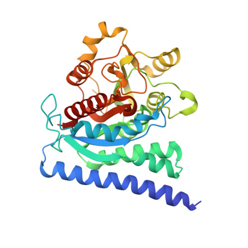
    Arginine acts as a precursor of polyamines in plants in two known pathways, agmatine and ornithine routes. It is decarboxylated to agmatine by arginine decarboxylase, and then transformed to putrescine by the consecutive action of agmatine iminohydrolase and N-carbamoylputrescine amidohydrolase. Alternatively, it can be hydrolyzed to ornithine by arginase and then decarboxylated by ornithine decarboxylase to putrescine. Some plants lack a functional ornithine pathway, but all have one or two arginases that can have dual cellular localization, in mitochondria and plastids. It was recently shown that arginases from Arabidopsis thaliana and soybean act also as agmatinases, thus they can produce putrescine directly from agmatine. Therefore, arginase (together with arginine decarboxylase) can complement putrescine production in plastids, providing a third polyamine biosynthesis pathway in plants. Phylogenetic analysis suggests that arginases, highly conserved in the plant kingdom, create the only group of enzymes recognized in the family of ureohydrolases in plants. Arginases are metalloenzymes with binuclear manganese cluster in the active site. In this work, two arginases from A. thaliana and Medicago truncatula are structurally characterized and their binding properties are discussed. Crystal structures with bound ornithine show that plant hexameric arginases engage a long loop from the neighboring subunit to stabilize α-amino and carboxyl groups of the ligand. This unique ligand binding mode is unobserved in arginases from other domains of life. Structural analysis shows that substrate binding by residues from two neighboring subunits might also characterize some prokaryotic agmatinases. This feature of plant arginases is most likely the determinant of their ability to recognize not only arginine but also agmatine as their substrates, thus, to act as arginase and agmatinase.


  • Organizational Affiliation
    • Synchrotron Radiation Research Section of Macromolecular Crystallography Laboratory, National Cancer Institute, Argonne, IL, United States.

Macromolecule Content 

  • Total Structure Weight: 846.68 kDa 
  • Atom Count: 63,508 
  • Modeled Residue Count: 7,593 
  • Deposited Residue Count: 7,704 
  • Unique protein chains: 1

Macromolecules

Find similar proteins by:|  3D Structure
Entity ID: 1
MoleculeChains  Sequence LengthOrganismDetailsImage
Arginase 1, mitochondrial
A, B, C, D, E
A, B, C, D, E, F, G, H, I, J, K, L, M, N, O, P, Q, R, S, T, U, V, W, X
321Arabidopsis thalianaMutation(s): 0 
Gene Names: ARGAH1At4g08900T3H13.7
EC: 3.5.3.1 (PDB Primary Data), 3.5.3.11 (PDB Primary Data)
UniProt
Find proteins for P46637 (Arabidopsis thaliana)
Explore P46637 
Go to UniProtKB:  P46637
Entity Groups
Sequence Clusters30% Identity50% Identity70% Identity90% Identity95% Identity100% Identity
UniProt GroupP46637
Sequence Annotations
Expand
Reference Sequence

Small Molecules

Ligands 5 Unique
IDChains Name / Formula / InChI Key2D Diagram3D Interactions
ORN
(Subject of Investigation/LOI)

Query on ORN


Download:Ideal Coordinates CCD File
BA [auth A]
BB [auth F]
BD [auth P]
BE [auth V]
EC [auth K]
BA [auth A],
BB [auth F],
BD [auth P],
BE [auth V],
EC [auth K],
FB [auth G],
LA [auth C],
UA [auth E],
XD [auth U]
L-ornithine
C5 H12 N2 O2
AHLPHDHHMVZTML-BYPYZUCNSA-N
PEG

Query on PEG


Download:Ideal Coordinates CCD File
HB [auth G],
IB [auth G],
KC [auth L],
LC [auth L],
MC [auth L]
DI(HYDROXYETHYL)ETHER
C4 H10 O3
MTHSVFCYNBDYFN-UHFFFAOYSA-N
GOL

Query on GOL


Download:Ideal Coordinates CCD File
AB [auth F]
BC [auth K]
DC [auth K]
EA [auth B]
ED [auth Q]
AB [auth F],
BC [auth K],
DC [auth K],
EA [auth B],
ED [auth Q],
EE [auth W],
GA [auth B],
GB [auth G],
GE [auth W],
HC [auth L],
JA [auth C],
JB [auth G],
JC [auth L],
JD [auth R],
MB [auth H],
MD [auth S],
NB [auth H],
OA [auth D],
OD [auth S],
QA [auth D],
SB [auth I],
SC [auth N],
SD [auth T],
TB [auth I],
VA [auth E],
VD [auth U],
WA [auth E],
XB [auth J],
YB [auth J],
ZC [auth P]
GLYCEROL
C3 H8 O3
PEDCQBHIVMGVHV-UHFFFAOYSA-N
MN
(Subject of Investigation/LOI)

Query on MN


Download:Ideal Coordinates CCD File
AC [auth K]
CA [auth B]
CB [auth G]
CD [auth Q]
CE [auth W]
AC [auth K],
CA [auth B],
CB [auth G],
CD [auth Q],
CE [auth W],
DA [auth B],
DB [auth G],
DD [auth Q],
DE [auth W],
FC [auth L],
GC [auth L],
GD [auth R],
HA [auth C],
HD [auth R],
HE [auth X],
IA [auth C],
IE [auth X],
KB [auth H],
KD [auth S],
LB [auth H],
LD [auth S],
MA [auth D],
NA [auth D],
NC [auth M],
OC [auth M],
PB [auth I],
PD [auth T],
QB [auth I],
QC [auth N],
QD [auth T],
RA [auth E],
RC [auth N],
SA [auth E],
TD [auth U],
UB [auth J],
UC [auth O],
UD [auth U],
VB [auth J],
VC [auth O],
XA [auth F],
XC [auth P],
Y [auth A],
YA [auth F],
YC [auth P],
YD [auth V],
Z [auth A],
ZB [auth K],
ZD [auth V]
MANGANESE (II) ION
Mn
WAEMQWOKJMHJLA-UHFFFAOYSA-N
NA

Query on NA


Download:Ideal Coordinates CCD File
AA [auth A]
AD [auth P]
AE [auth V]
CC [auth K]
EB [auth G]
AA [auth A],
AD [auth P],
AE [auth V],
CC [auth K],
EB [auth G],
FA [auth B],
FD [auth Q],
FE [auth W],
IC [auth L],
ID [auth R],
JE [auth X],
KA [auth C],
ND [auth S],
OB [auth H],
PA [auth D],
PC [auth M],
RB [auth I],
RD [auth T],
TA [auth E],
TC [auth N],
WB [auth J],
WC [auth O],
WD [auth U],
ZA [auth F]
SODIUM ION
Na
FKNQFGJONOIPTF-UHFFFAOYSA-N

Experimental Data & Validation

Experimental Data

  • Method: X-RAY DIFFRACTION
  • Resolution: 2.25 Å
  • R-Value Free:  0.197 (Depositor), 0.202 (DCC) 
  • R-Value Work:  0.159 (Depositor), 0.167 (DCC) 
  • R-Value Observed: 0.159 (Depositor) 
Space Group: I 41
Unit Cell:
Length ( Å )Angle ( ˚ )
a = 267.322α = 90
b = 267.322β = 90
c = 262.894γ = 90
Software Package:
Software NamePurpose
REFMACrefinement
PDB_EXTRACTdata extraction
HKL-3000data reduction
HKL-3000data scaling
PHASERphasing

Structure Validation

View Full Validation Report


Ligand Structure Quality Assessment 


Entry History 

& Funding Information

Deposition Data

  • Released Date: 2020-08-12 
  • Deposition Author(s): Sekula, B.

Funding OrganizationLocationGrant Number
National Institutes of Health/National Cancer Institute (NIH/NCI)United StatesIntramural Research Program

Revision History  (Full details and data files)

  • Version 1.0: 2020-08-12
    Type: Initial release
  • Version 1.1: 2020-08-19
    Changes: Database references
  • Version 1.2: 2023-10-11
    Changes: Data collection, Database references, Refinement description