6VQ5

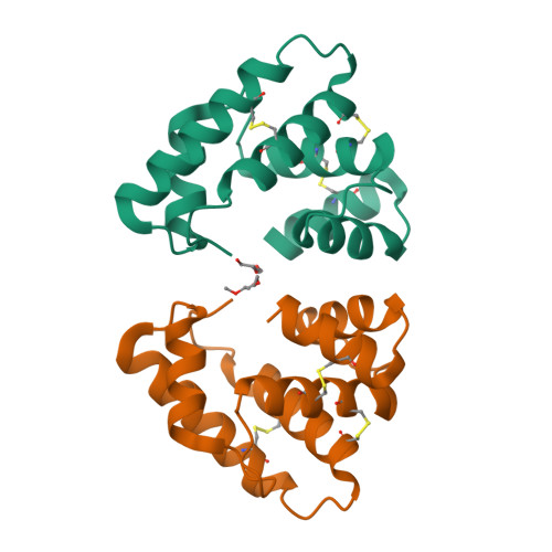
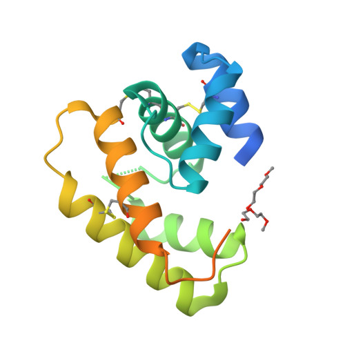
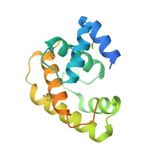
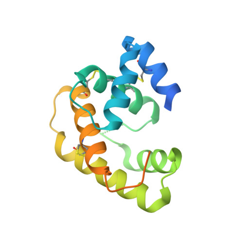
Crystal Structure of Epiphyas postvittana Pheromone Binding Protein 3


Experimental Data Snapshot

  • Method: X-RAY DIFFRACTION
  • Resolution: 2.60 Å
  • R-Value Free: 0.259 
  • R-Value Work: 0.216 
  • R-Value Observed: 0.218 

Starting Model: experimental
View more details

wwPDB Validation   3D Report Full Report

Currently 6VQ5 does not have a validation slider image.


This is version 1.2 of the entry. See complete history


Literature

Crystal structure of Epiphyas postvittana pheromone binding protein 3.

Hamiaux, C.Carraher, C.Lofstedt, C.Corcoran, J.A.

(2020) Sci Rep 10: 16366-16366

  • DOI: https://doi.org/10.1038/s41598-020-73294-8
  • Primary Citation of Related Structures:  
    6VQ5

  • PubMed Abstract: 

    The insect olfactory system operates as a well-choreographed ensemble of molecules which functions to selectively translate volatile chemical messages present in the environment into neuronal impulses that guide insect behaviour. Of these molecules, binding proteins are believed to transport hydrophobic odorant molecules across the aqueous lymph present in antennal sensilla to receptors present in olfactory sensory neurons. Though the exact mechanism through which these proteins operate is still under investigation, these carriers clearly play a critical role in determining what an insect can smell. Binding proteins that transport important sex pheromones are colloquially named pheromone binding proteins (PBPs). Here, we have produced a functional recombinant PBP from the horticultural pest, Epiphyas postvittana (EposPBP3), and experimentally solved its apo-structure through X-ray crystallography to a resolution of 2.60 Å. Structural comparisons with related lepidopteran PBPs further allowed us to propose models for the binding of pheromone components to EposPBP3. The data presented here represent the first structure of an olfactory-related protein from the tortricid family of moths, whose members cause billions of dollars in losses to agricultural producers each year. Knowledge of the structure of these important proteins will allow for subsequent studies in which novel, olfactory molecule-specific insecticides can be developed.


  • Organizational Affiliation

    The New Zealand Institute for Plant and Food Research Limited, Auckland, New Zealand.


Macromolecules
Find similar proteins by:  (by identity cutoff)  |  3D Structure
Entity ID: 1
MoleculeChains Sequence LengthOrganismDetailsImage
Pheromome Binding Protein
A, B
159Epiphyas postvittanaMutation(s): 1 
UniProt
Find proteins for A0A0K8TU48 (Epiphyas postvittana)
Explore A0A0K8TU48 
Go to UniProtKB:  A0A0K8TU48
Entity Groups  
Sequence Clusters30% Identity50% Identity70% Identity90% Identity95% Identity100% Identity
UniProt GroupA0A0K8TU48
Sequence Annotations
Expand
  • Reference Sequence
Small Molecules
Ligands 1 Unique
IDChains Name / Formula / InChI Key2D Diagram3D Interactions
PG0
Query on PG0

Download Ideal Coordinates CCD File 
C [auth A],
D [auth A]
2-(2-METHOXYETHOXY)ETHANOL
C5 H12 O3
SBASXUCJHJRPEV-UHFFFAOYSA-N
Experimental Data & Validation

Experimental Data

  • Method: X-RAY DIFFRACTION
  • Resolution: 2.60 Å
  • R-Value Free: 0.259 
  • R-Value Work: 0.216 
  • R-Value Observed: 0.218 
  • Space Group: P 43
Unit Cell:
Length ( Å )Angle ( ˚ )
a = 53.379α = 90
b = 53.379β = 90
c = 105.949γ = 90
Software Package:
Software NamePurpose
XDSdata reduction
Aimlessdata scaling
REFMACrefinement
PDB_EXTRACTdata extraction
MoRDaphasing

Structure Validation

View Full Validation Report

Currently 6VQ5 does not have a validation slider image.


Entry History 

Deposition Data

Revision History  (Full details and data files)

  • Version 1.0: 2020-10-14
    Type: Initial release
  • Version 1.1: 2023-10-11
    Changes: Data collection, Database references, Refinement description
  • Version 1.2: 2024-10-23
    Changes: Structure summary