6VLY | pdb_00006vly

Crystal structure of FabI-NADH complex from Alistipes finegoldii


Experimental Data Snapshot

  • Method: X-RAY DIFFRACTION
  • Resolution: 1.86 Å
  • R-Value Free: 
    0.197 (Depositor), 0.205 (DCC) 
  • R-Value Work: 
    0.158 (Depositor), 0.170 (DCC) 
  • R-Value Observed: 
    0.160 (Depositor) 

Starting Model: experimental
View more details

wwPDB Validation 3D Report Full Report

Validation slider image for 6VLY

Ligand Structure Quality Assessment 


This is version 1.3 of the entry. See complete history

Literature

The genome of a Bacteroidetes inhabitant of the human gut encodes a structurally distinct enoyl-acyl carrier protein reductase (FabI).

Radka, C.D.Frank, M.W.Yao, J.Seetharaman, J.Miller, D.J.Rock, C.O.

(2020) J Biological Chem 295: 7635-7652

  • DOI: https://doi.org/10.1074/jbc.RA120.013336
  • Primary Citation Related Structures: 
    6VLX, 6VLY

  • PubMed Abstract: 

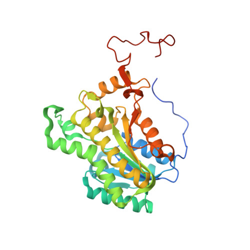
    Enoyl-acyl carrier protein reductase (FabI) catalyzes a rate-controlling step in bacterial fatty-acid synthesis and is a target for antibacterial drug development. A phylogenetic analysis shows that FabIs fall into four divergent clades. Members of clades 1-3 have been structurally and biochemically characterized, but the fourth clade, found in members of phylum Bacteroidetes, is uncharacterized. Here, we identified the unique structure and conformational changes that distinguish clade 4 FabIs. Alistipes finegoldii is a prototypical Bacteroidetes inhabitant of the gut microbiome. We found that A. finegoldii FabI ( Af FabI) displays cooperative kinetics and uses NADH as a cofactor, and its crystal structure at 1.72 Å resolution showed that it adopts a Rossmann fold as do other characterized FabIs. It also disclosed a carboxyl-terminal extension that forms a helix-helix interaction that links the protomers as a unique feature of Af FabI. An A f FabI·NADH crystal structure at 1.86 Å resolution revealed that this feature undergoes a large conformational change to participate in covering the NADH-binding pocket and establishing the water channels that connect the active site to the central water well. Progressive deletion of these interactions led to catalytically compromised proteins that fail to bind NADH. This unique conformational change imparted a distinct shape to the Af FabI active site that renders it refractory to a FabI drug that targets clade 1 and 3 pathogens. We conclude that the clade 4 FabI, found in the Bacteroidetes inhabitants of the gut, have several structural features and conformational transitions that distinguish them from other bacterial FabIs.


  • Organizational Affiliation
    • Department of Infectious Diseases, St. Jude Children's Research Hospital, Memphis, Tennessee 38105.

Macromolecule Content 

  • Total Structure Weight: 139.96 kDa 
  • Atom Count: 10,132 
  • Modeled Residue Count: 1,144 
  • Deposited Residue Count: 1,236 
  • Unique protein chains: 1

Macromolecules

Find similar proteins by:|  3D Structure
Entity ID: 1
MoleculeChains  Sequence LengthOrganismDetailsImage
Enoyl-[acyl-carrier-protein] reductase [NADH]
A, B, C, D
309Alistipes finegoldiiMutation(s): 0 
Gene Names: fabIERS852447_01935
EC: 1.3.1.9

Small Molecules

Ligands 3 Unique
IDChains Name / Formula / InChI Key2D Diagram3D Interactions
NAI
(Subject of Investigation/LOI)

Query on NAI


Download:Ideal Coordinates CCD File
F [auth A],
J [auth B],
N [auth C],
Q [auth D]
1,4-DIHYDRONICOTINAMIDE ADENINE DINUCLEOTIDE
C21 H29 N7 O14 P2
BOPGDPNILDQYTO-NNYOXOHSSA-N
GOL

Query on GOL


Download:Ideal Coordinates CCD File
E [auth A]
G [auth B]
H [auth B]
I [auth B]
L [auth C]
E [auth A],
G [auth B],
H [auth B],
I [auth B],
L [auth C],
M [auth C],
O [auth D],
P [auth D]
GLYCEROL
C3 H8 O3
PEDCQBHIVMGVHV-UHFFFAOYSA-N
CL

Query on CL


Download:Ideal Coordinates CCD File
K [auth B]CHLORIDE ION
Cl
VEXZGXHMUGYJMC-UHFFFAOYSA-M

Experimental Data & Validation

Experimental Data

  • Method: X-RAY DIFFRACTION
  • Resolution: 1.86 Å
  • R-Value Free:  0.197 (Depositor), 0.205 (DCC) 
  • R-Value Work:  0.158 (Depositor), 0.170 (DCC) 
  • R-Value Observed: 0.160 (Depositor) 
Space Group: P 21 21 21
Unit Cell:
Length ( Å )Angle ( ˚ )
a = 75.313α = 90
b = 111.537β = 90
c = 150.54γ = 90
Software Package:
Software NamePurpose
REFMACrefinement
PDB_EXTRACTdata extraction
XDSdata reduction
Aimlessdata scaling
PHASERphasing

Structure Validation

View Full Validation Report


Ligand Structure Quality Assessment 


Entry History 

& Funding Information

Deposition Data


Funding OrganizationLocationGrant Number
National Institutes of Health/National Institute of General Medical Sciences (NIH/NIGMS)United StatesGM034496
National Institutes of Health/National Cancer Institute (NIH/NCI)United StatesCA21765

Revision History  (Full details and data files)

  • Version 1.0: 2020-04-29
    Type: Initial release
  • Version 1.1: 2020-05-06
    Changes: Database references
  • Version 1.2: 2020-06-10
    Changes: Database references
  • Version 1.3: 2023-10-11
    Changes: Data collection, Database references, Refinement description