6VK3 | pdb_00006vk3

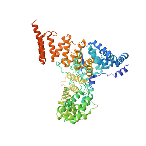
CryoEM structure of Hrd3/Yos9 complex


Experimental Data Snapshot

  • Method: ELECTRON MICROSCOPY
  • Resolution: 3.70 Å
  • Aggregation State: PARTICLE 
  • Reconstruction Method: SINGLE PARTICLE 

wwPDB Validation 3D Report Full Report

Validation slider image for 6VK3

This is version 1.2 of the entry. See complete history. 

Literature

Structural basis of ER-associated protein degradation mediated by the Hrd1 ubiquitin ligase complex.

Wu, X., Siggel, M., Ovchinnikov, S., Mi, W., Svetlov, V., Nudler, E., Liao, M., Hummer, G., Rapoport, T.A.

(2020) Science 368

  • DOI: https://doi.org/10.1126/science.aaz2449
  • Primary Citation Related Structures: 
    6VJY, 6VJZ, 6VK0, 6VK1, 6VK3

  • PubMed Abstract: 

    Misfolded luminal endoplasmic reticulum (ER) proteins undergo ER-associated degradation (ERAD-L): They are retrotranslocated into the cytosol, polyubiquitinated, and degraded by the proteasome. ERAD-L is mediated by the Hrd1 complex (composed of Hrd1, Hrd3, Der1, Usa1, and Yos9), but the mechanism of retrotranslocation remains mysterious. Here, we report a structure of the active Hrd1 complex, as determined by cryo-electron microscopy analysis of two subcomplexes. Hrd3 and Yos9 jointly create a luminal binding site that recognizes glycosylated substrates. Hrd1 and the rhomboid-like Der1 protein form two "half-channels" with cytosolic and luminal cavities, respectively, and lateral gates facing one another in a thinned membrane region. These structures, along with crosslinking and molecular dynamics simulation results, suggest how a polypeptide loop of an ERAD-L substrate moves through the ER membrane.


  • Organizational Affiliation: 
    • Howard Hughes Medical Institute and Department of Cell Biology, Harvard Medical School, Boston, MA 02115, USA.

Macromolecule Content 

  • Total Structure Weight: 145.64 kDa 
  • Atom Count: 7,784 
  • Modeled Residue Count: 961 
  • Deposited Residue Count: 1,274 
  • Unique protein chains: 2

Macromolecules

Find similar proteins by:|  3D Structure
Entity ID: 1
MoleculeChains  Sequence LengthOrganismDetailsImage
Hrd3732Saccharomyces cerevisiaeMutation(s): 0 
Gene Names: Hrd3
Membrane Entity: Yes 
UniProt
Find proteins for Q05787 (Saccharomyces cerevisiae (strain ATCC 204508 / S288c))
Explore Q05787 
Go to UniProtKB:  Q05787
Entity Groups
Sequence Clusters30% Identity50% Identity70% Identity90% Identity95% Identity100% Identity
UniProt GroupQ05787
Sequence Annotations
Expand
Reference Sequence
Find similar proteins by:|  3D Structure
Entity ID: 2
MoleculeChains  Sequence LengthOrganismDetailsImage
Protein OS-9 homolog542Saccharomyces cerevisiaeMutation(s): 0 
Gene Names: YOS9
Membrane Entity: Yes 
UniProt
Find proteins for Q99220 (Saccharomyces cerevisiae (strain ATCC 204508 / S288c))
Explore Q99220 
Go to UniProtKB:  Q99220
Entity Groups
Sequence Clusters30% Identity50% Identity70% Identity90% Identity95% Identity100% Identity
UniProt GroupQ99220
Sequence Annotations
Expand
Reference Sequence

Small Molecules

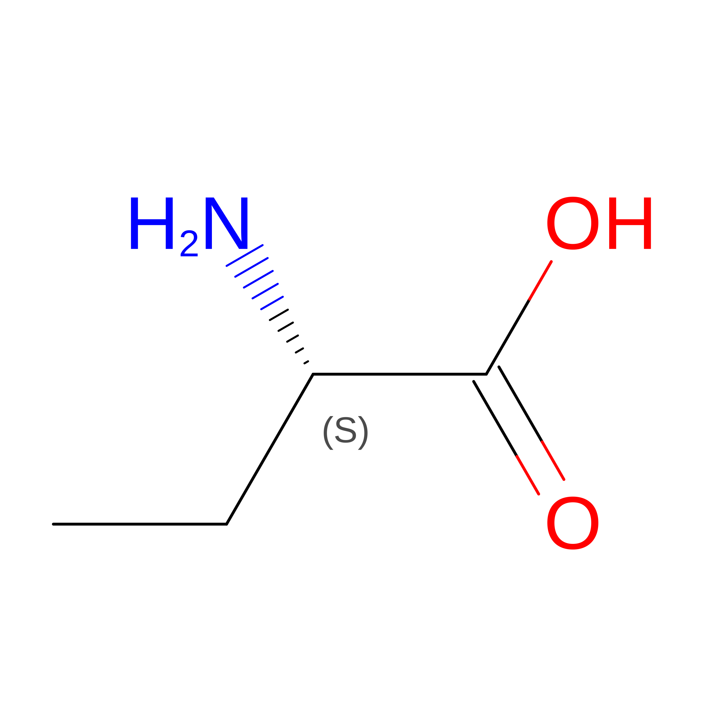
Modified Residues  1 Unique
IDChains TypeFormula2D DiagramParent
UNK
Query on UNK
A
L-PEPTIDE LINKINGC4 H9 N O2

--

Experimental Data & Validation

Experimental Data

  • Method: ELECTRON MICROSCOPY
  • Resolution: 3.70 Å
  • Aggregation State: PARTICLE 
  • Reconstruction Method: SINGLE PARTICLE 
EM Software:
TaskSoftware PackageVersion
RECONSTRUCTIONRELION3.0
MODEL REFINEMENTPHENIX

Structure Validation

View Full Validation Report


Entry History 

& Funding Information

Deposition Data


Funding OrganizationLocationGrant Number
National Institutes of Health/National Institute of General Medical Sciences (NIH/NIGMS)United StatesR01GM052586

Revision History  (Full details and data files)

  • Version 1.0: 2020-04-29
    Type: Initial release
  • Version 1.1: 2020-05-06
    Changes: Database references
  • Version 1.2: 2024-10-23
    Changes: Data collection, Database references, Structure summary