6VHY | pdb_00006vhy

NpsA-ThdA, an artificially fused Adenylation-PCP di-domain NRPS from Klebsiella oxytoca


Experimental Data Snapshot

  • Method: X-RAY DIFFRACTION
  • Resolution: 3.00 Å
  • R-Value Free: 
    0.260 (Depositor), 0.263 (DCC) 
  • R-Value Work: 
    0.214 (Depositor), 0.217 (DCC) 
  • R-Value Observed: 
    0.216 (Depositor) 

Starting Model: experimental
View more details

wwPDB Validation 3D Report Full Report

Validation slider image for 6VHY

Ligand Structure Quality Assessment 


This is version 1.3 of the entry. See complete history

Literature

Biosynthesis, Mechanism of Action, and Inhibition of the Enterotoxin Tilimycin Produced by the Opportunistic PathogenKlebsiella oxytoca.

Alexander, E.M.Kreitler, D.F.Guidolin, V.Hurben, A.K.Drake, E.Villalta, P.W.Balbo, S.Gulick, A.M.Aldrich, C.C.

(2020) ACS Infect Dis 6: 1976-1997

  • DOI: https://doi.org/10.1021/acsinfecdis.0c00326
  • Primary Citation Related Structures: 
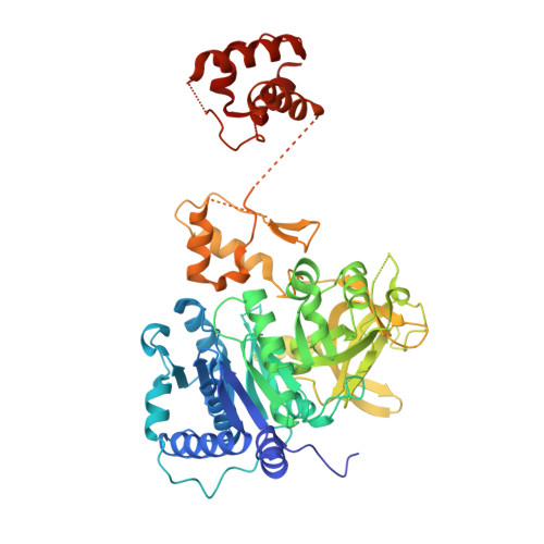
    6VHT, 6VHU, 6VHV, 6VHW, 6VHX, 6VHY, 6VHZ

  • PubMed Abstract: 

    Tilimycin is an enterotoxin produced by the opportunistic pathogen Klebsiella oxytoca that causes antibiotic-associated hemorrhagic colitis (AAHC). This pyrrolobenzodiazepine (PBD) natural product is synthesized by a bimodular nonribosomal peptide synthetase (NRPS) pathway composed of three proteins: NpsA, ThdA, and NpsB. We describe the functional and structural characterization of the fully reconstituted NRPS system and report the steady-state kinetic analysis of all natural substrates and cofactors as well as the structural characterization of both NpsA and ThdA. The mechanism of action of tilimycin was confirmed using DNA adductomics techniques through the detection of putative N-2 guanine alkylation after tilimycin exposure to eukaryotic cells, providing the first structural characterization of a PBD-DNA adduct formed in cells. Finally, we report the rational design of small-molecule inhibitors that block tilimycin biosynthesis in whole cell K. oxytoca (IC 50 = 29 ± 4 μM) through the inhibition of NpsA ( K D = 29 ± 4 nM).


  • Organizational Affiliation
    • Department of Medicinal Chemistry, University of Minnesota, Minneapolis, Minnesota 55455, United States.

Macromolecule Content 

  • Total Structure Weight: 259.48 kDa 
  • Atom Count: 15,436 
  • Modeled Residue Count: 2,093 
  • Deposited Residue Count: 2,344 
  • Unique protein chains: 1

Macromolecules

Find similar proteins by:|  3D Structure
Entity ID: 1
MoleculeChains  Sequence LengthOrganismDetailsImage
NpsA Adenylation Domain, Non-ribosomal peptide synthetase fusion protein
A, B, C, D
586Klebsiella oxytocaMutation(s): 4 
Gene Names: AGF18_11095AGF18_11090

Experimental Data & Validation

Experimental Data

  • Method: X-RAY DIFFRACTION
  • Resolution: 3.00 Å
  • R-Value Free:  0.260 (Depositor), 0.263 (DCC) 
  • R-Value Work:  0.214 (Depositor), 0.217 (DCC) 
  • R-Value Observed: 0.216 (Depositor) 
Space Group: P 1
Unit Cell:
Length ( Å )Angle ( ˚ )
a = 58.54α = 95.331
b = 98.721β = 96.337
c = 118.754γ = 106.508
Software Package:
Software NamePurpose
PHENIXrefinement
XDSdata reduction
Aimlessdata scaling
autoPROCdata scaling
PHASERphasing
Cootmodel building

Structure Validation

View Full Validation Report


Ligand Structure Quality Assessment 


Entry History 

& Funding Information

Deposition Data


Funding OrganizationLocationGrant Number
National Institutes of Health/National Institute of General Medical Sciences (NIH/NIGMS)United StatesGM116957

Revision History  (Full details and data files)

  • Version 1.0: 2020-06-24
    Type: Initial release
  • Version 1.1: 2020-07-22
    Changes: Database references
  • Version 1.2: 2023-10-11
    Changes: Data collection, Database references, Refinement description
  • Version 1.3: 2024-11-20
    Changes: Structure summary