6VHH | pdb_00006vhh

Human Teneurin-2 and human Latrophilin-3 binary complex


Experimental Data Snapshot

  • Method: ELECTRON MICROSCOPY
  • Resolution: 2.97 Å
  • Aggregation State: PARTICLE 
  • Reconstruction Method: SINGLE PARTICLE 

wwPDB Validation 3D Report Full Report

Validation slider image for 6VHH

This is version 2.1 of the entry. See complete history

Literature

Alternative splicing controls teneurin-latrophilin interaction and synapse specificity by a shape-shifting mechanism.

Li, J.Xie, Y.Cornelius, S.Jiang, X.Sando, R.Kordon, S.P.Pan, M.Leon, K.Sudhof, T.C.Zhao, M.Arac, D.

(2020) Nat Commun 11: 2140-2140

  • DOI: https://doi.org/10.1038/s41467-020-16029-7
  • Primary Citation Related Structures: 
    6VHH

  • PubMed Abstract: 

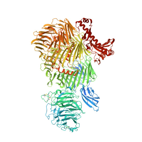
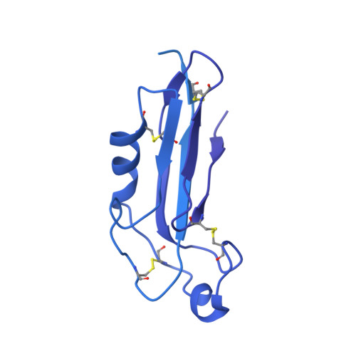
    The trans-synaptic interaction of the cell-adhesion molecules teneurins (TENs) with latrophilins (LPHNs/ADGRLs) promotes excitatory synapse formation when LPHNs simultaneously interact with FLRTs. Insertion of a short alternatively-spliced region within TENs abolishes the TEN-LPHN interaction and switches TEN function to specify inhibitory synapses. How alternative-splicing regulates TEN-LPHN interaction remains unclear. Here, we report the 2.9 Å resolution cryo-EM structure of the TEN2-LPHN3 complex, and describe the trimeric TEN2-LPHN3-FLRT3 complex. The structure reveals that the N-terminal lectin domain of LPHN3 binds to the TEN2 barrel at a site far away from the alternatively spliced region. Alternative-splicing regulates the TEN2-LPHN3 interaction by hindering access to the LPHN-binding surface rather than altering it. Strikingly, mutagenesis of the LPHN-binding surface of TEN2 abolishes the LPHN3 interaction and impairs excitatory but not inhibitory synapse formation. These results suggest that a multi-level coincident binding mechanism mediated by a cryptic adhesion complex between TENs and LPHNs regulates synapse specificity.


  • Organizational Affiliation
    • Department of Biochemistry and Molecular Biology, The University of Chicago, Chicago, IL, 60637, USA.

Macromolecule Content 

  • Total Structure Weight: 314.57 kDa 
  • Atom Count: 14,219 
  • Modeled Residue Count: 1,821 
  • Deposited Residue Count: 2,780 
  • Unique protein chains: 2

Macromolecules

Find similar proteins by:|  3D Structure
Entity ID: 1
MoleculeChains  Sequence LengthOrganismDetailsImage
Teneurin-21,928Homo sapiensMutation(s): 0 
Gene Names: TENM2KIAA1127ODZ2TNM2
UniProt & NIH Common Fund Data Resources
Find proteins for Q9NT68 (Homo sapiens)
Explore Q9NT68 
Go to UniProtKB:  Q9NT68
PHAROS:  Q9NT68
GTEx:  ENSG00000145934 
Entity Groups
Sequence Clusters30% Identity50% Identity70% Identity90% Identity95% Identity100% Identity
UniProt GroupQ9NT68
Glycosylation
Glycosylation Sites: 8Go to GlyGen: Q9NT68-1
Sequence Annotations
Expand
Reference Sequence
Find similar proteins by:|  3D Structure
Entity ID: 2
MoleculeChains  Sequence LengthOrganismDetailsImage
Adhesion G protein-coupled receptor L3852Homo sapiensMutation(s): 0 
Gene Names: ADGRL3KIAA0768LEC3LPHN3
UniProt & NIH Common Fund Data Resources
Find proteins for Q9HAR2 (Homo sapiens)
Explore Q9HAR2 
Go to UniProtKB:  Q9HAR2
PHAROS:  Q9HAR2
GTEx:  ENSG00000150471 
Entity Groups
Sequence Clusters30% Identity50% Identity70% Identity90% Identity95% Identity100% Identity
UniProt GroupQ9HAR2
Sequence Annotations
Expand
Reference Sequence

Oligosaccharides

Help  
Entity ID: 3
MoleculeChains Length2D Diagram GlycosylationD Interactions
alpha-D-mannopyranose-(1-4)-2-acetamido-2-deoxy-beta-D-glucopyranose-(1-4)-2-acetamido-2-deoxy-beta-D-glucopyranose
C
3N-Glycosylation
Glycosylation Resources
GlyTouCan: G62182OO
GlyCosmos: G62182OO
GlyGen: G62182OO
Entity ID: 4
MoleculeChains Length2D Diagram GlycosylationD Interactions
beta-D-mannopyranose-(1-4)-2-acetamido-2-deoxy-beta-D-glucopyranose-(1-4)-2-acetamido-2-deoxy-beta-D-glucopyranose
D
3N-Glycosylation
Glycosylation Resources
GlyTouCan: G15407YE
GlyCosmos: G15407YE
GlyGen: G15407YE
Entity ID: 5
MoleculeChains Length2D Diagram GlycosylationD Interactions
2-acetamido-2-deoxy-beta-D-glucopyranose-(1-4)-2-acetamido-2-deoxy-beta-D-glucopyranose
E, F, G
2N-Glycosylation
Glycosylation Resources
GlyTouCan: G42666HT
GlyCosmos: G42666HT
GlyGen: G42666HT

Experimental Data & Validation

Experimental Data

  • Method: ELECTRON MICROSCOPY
  • Resolution: 2.97 Å
  • Aggregation State: PARTICLE 
  • Reconstruction Method: SINGLE PARTICLE 
EM Software:
TaskSoftware PackageVersion
RECONSTRUCTIONRELION3.0.8

Structure Validation

View Full Validation Report


Entry History 

& Funding Information

Deposition Data


Funding OrganizationLocationGrant Number
National Institutes of Health/National Institute of General Medical Sciences (NIH/NIGMS)United StatesR01 GM120322
National Institutes of Health/National Institute of General Medical Sciences (NIH/NIGMS)United StatesR01 GM134035-01
American Heart AssociationUnited States19POST34380439/Jingxian Li/2019-2020

Revision History  (Full details and data files)

  • Version 1.0: 2020-06-03
    Type: Initial release
  • Version 2.0: 2020-07-29
    Type: Remediation
    Reason: Carbohydrate remediation
    Changes: Advisory, Atomic model, Data collection, Derived calculations, Structure summary
  • Version 2.1: 2024-10-23
    Changes: Data collection, Database references, Structure summary