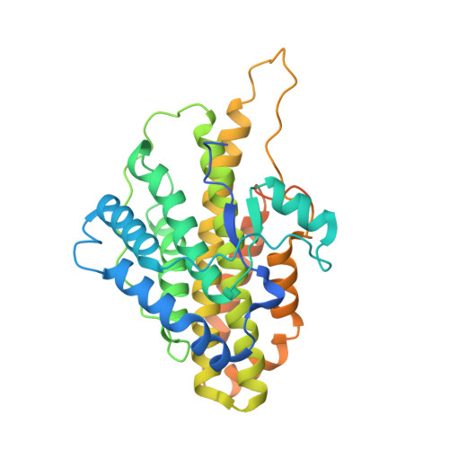
6VE0

Crystal structure of reduced SfmD by soaking with sodium hydrosulfite


Experimental Data Snapshot

  • Method: X-RAY DIFFRACTION
  • Resolution: 3.15 Å
  • R-Value Free: 0.308 
  • R-Value Work: 0.260 
  • R-Value Observed: 0.265 

Starting Model: experimental
View more details

wwPDB Validation   3D Report Full Report

Currently 6VE0 does not have a validation slider image.


This is version 1.3 of the entry. See complete history


Literature

A novel catalytic heme cofactor in SfmD with a single thioether bond and a bis -His ligand set revealed by a de novo crystal structural and spectroscopic study.

Shin, I.Davis, I.Nieves-Merced, K.Wang, Y.McHardy, S.Liu, A.

(2021) Chem Sci 12: 3984-3998

  • DOI: https://doi.org/10.1039/d0sc06369j
  • Primary Citation of Related Structures:  
    6VDP, 6VDQ, 6VDZ, 6VE0

  • PubMed Abstract: 

    SfmD is a heme-dependent enzyme in the biosynthetic pathway of saframycin A. Here, we present a 1.78 Å resolution de novo crystal structure of SfmD, which unveils a novel heme cofactor attached to the protein with an unusual H x n H xxx C motif ( n ∼ 38). This heme cofactor is unique in two respects. It contains a single thioether bond in a cysteine-vinyl link with Cys317, and the ferric heme has two axial protein ligands, i.e. , His274 and His313. We demonstrated that SfmD heme is catalytically active and can utilize dioxygen and ascorbate for a single-oxygen insertion into 3-methyl-l-tyrosine. Catalytic assays using ascorbate derivatives revealed the functional groups of ascorbate essential to its function as a cosubstrate. Abolishing the thioether linkage through mutation of Cys317 resulted in catalytically inactive SfmD variants. EPR and optical data revealed that the heme center undergoes a substantial conformational change with one axial histidine ligand dissociating from the iron ion in response to substrate 3-methyl-l-tyrosine binding or chemical reduction by a reducing agent, such as the cosubstrate ascorbate. The labile axial ligand was identified as His274 through redox-linked structural determinations. Together, identifying an unusual heme cofactor with a previously unknown heme-binding motif for a monooxygenase activity and the structural similarity of SfmD to the members of the heme-based tryptophan dioxygenase superfamily will broaden understanding of heme chemistry.


  • Organizational Affiliation

    Department of Chemistry, The University of Texas at San Antonio One UTSA Circle Texas 78249 USA Feradical@utsa.edu.


Macromolecules
Find similar proteins by:  (by identity cutoff)  |  3D Structure
Entity ID: 1
MoleculeChains Sequence LengthOrganismDetailsImage
3-methyl-L-tyrosine peroxygenase365Streptomyces lavendulaeMutation(s): 0 
Gene Names: sfmD
EC: 1.11.2.5
UniProt
Find proteins for B0CN28 (Streptomyces lavendulae)
Explore B0CN28 
Go to UniProtKB:  B0CN28
Entity Groups  
Sequence Clusters30% Identity50% Identity70% Identity90% Identity95% Identity100% Identity
UniProt GroupB0CN28
Sequence Annotations
Expand
  • Reference Sequence
Small Molecules
Ligands 1 Unique
IDChains Name / Formula / InChI Key2D Diagram3D Interactions
HEC (Subject of Investigation/LOI)
Query on HEC

Download Ideal Coordinates CCD File 
B [auth A]HEME C
C34 H34 Fe N4 O4
HXQIYSLZKNYNMH-LJNAALQVSA-N
Experimental Data & Validation

Experimental Data

  • Method: X-RAY DIFFRACTION
  • Resolution: 3.15 Å
  • R-Value Free: 0.308 
  • R-Value Work: 0.260 
  • R-Value Observed: 0.265 
  • Space Group: P 31 2 1
Unit Cell:
Length ( Å )Angle ( ˚ )
a = 59.453α = 90
b = 59.453β = 90
c = 159.965γ = 120
Software Package:
Software NamePurpose
DENZOdata reduction
SCALEPACKdata scaling
PHENIXrefinement
PDB_EXTRACTdata extraction
PHENIXphasing

Structure Validation

View Full Validation Report

Currently 6VE0 does not have a validation slider image.


Entry History & Funding Information

Deposition Data


Funding OrganizationLocationGrant Number
National Institutes of Health/National Institute of General Medical Sciences (NIH/NIGMS)United States--

Revision History  (Full details and data files)

  • Version 1.0: 2021-03-10
    Type: Initial release
  • Version 1.1: 2021-07-14
    Changes: Database references
  • Version 1.2: 2023-10-11
    Changes: Data collection, Database references, Refinement description
  • Version 1.3: 2024-10-09
    Changes: Structure summary