6VAT | pdb_00006vat

Structure of the periplasmic domain of YejM from Salmonella typhimurium


Experimental Data Snapshot

  • Method: X-RAY DIFFRACTION
  • Resolution: 2.35 Å
  • R-Value Free: 
    0.249 (Depositor), 0.249 (DCC) 
  • R-Value Work: 
    0.196 (Depositor), 0.197 (DCC) 
  • R-Value Observed: 
    0.199 (Depositor) 

Starting Model: experimental
View more details

wwPDB Validation 3D Report Full Report

Validation slider image for 6VAT

This is version 2.1 of the entry. See complete history

Literature

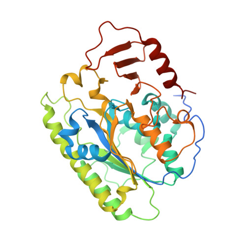
The essential inner membrane protein YejM is a metalloenzyme.

Gabale, U.Pena Palomino, P.A.Kim, H.Chen, W.Ressl, S.

(2020) Sci Rep 10: 17794-17794

  • DOI: https://doi.org/10.1038/s41598-020-73660-6
  • Primary Citation Related Structures: 
    6VAT, 6VC7, 6VDF

  • PubMed Abstract: 

    Recent recurrent outbreaks of Gram-negative bacteria show the critical need to target essential bacterial mechanisms to fight the increase of antibiotic resistance. Pathogenic Gram-negative bacteria have developed several strategies to protect themselves against the host immune response and antibiotics. One such strategy is to remodel the outer membrane where several genes are involved. yejM was discovered as an essential gene in E. coli and S. typhimurium that plays a critical role in their virulence by changing the outer membrane permeability. How the inner membrane protein YejM with its periplasmic domain changes membrane properties remains unknown. Despite overwhelming structural similarity between the periplasmic domains of two YejM homologues with hydrolases like arylsulfatases, no enzymatic activity has been previously reported for YejM. Our studies reveal an intact active site with bound metal ions in the structure of YejM periplasmic domain. Furthermore, we show that YejM has a phosphatase activity that is dependent on the presence of magnesium ions and is linked to its function of regulating outer membrane properties. Understanding the molecular mechanism by which YejM is involved in outer membrane remodeling will help to identify a new drug target in the fight against the increased antibiotic resistance.


  • Organizational Affiliation
    • Department of Molecular and Cellular Biochemistry, Indiana University Bloomington, 212 S Hawthrone Dr, Bloomington, IN, 47405, USA. ugabale@indiana.edu.

Macromolecule Content 

  • Total Structure Weight: 250.83 kDa 
  • Atom Count: 17,290 
  • Modeled Residue Count: 2,066 
  • Deposited Residue Count: 2,208 
  • Unique protein chains: 1

Macromolecules

Find similar proteins by:|  3D Structure
Entity ID: 1
MoleculeChains  Sequence LengthOrganismDetailsImage
Periplasmic domain of the cardiolipin transporter protein YejM/PbgA
A, B, C, D, E
A, B, C, D, E, F
368Salmonella enterica subsp. enterica serovar 1,4,[5],12:i:-Mutation(s): 0 
Gene Names: pbgAFGF59_12130
EC: 3.1
UniProt
Find proteins for P40709 (Salmonella typhimurium (strain LT2 / SGSC1412 / ATCC 700720))
Explore P40709 
Go to UniProtKB:  P40709
Entity Groups
Sequence Clusters30% Identity50% Identity70% Identity90% Identity95% Identity100% Identity
UniProt GroupP40709
Sequence Annotations
Expand
Reference Sequence

Small Molecules

Ligands 8 Unique
IDChains Name / Formula / InChI Key2D Diagram3D Interactions
P6G

Query on P6G


Download:Ideal Coordinates CCD File
BA [auth D]HEXAETHYLENE GLYCOL
C12 H26 O7
IIRDTKBZINWQAW-UHFFFAOYSA-N
1PE

Query on 1PE


Download:Ideal Coordinates CCD File
EA [auth E],
HA [auth F],
K [auth A],
V [auth C]
PENTAETHYLENE GLYCOL
C10 H22 O6
JLFNLZLINWHATN-UHFFFAOYSA-N
PG4

Query on PG4


Download:Ideal Coordinates CCD File
DA [auth E]
G [auth A]
H [auth A]
M [auth B]
Q [auth C]
DA [auth E],
G [auth A],
H [auth A],
M [auth B],
Q [auth C],
R [auth C],
Y [auth D]
TETRAETHYLENE GLYCOL
C8 H18 O5
UWHCKJMYHZGTIT-UHFFFAOYSA-N
PGE

Query on PGE


Download:Ideal Coordinates CCD File
GA [auth F],
N [auth B],
S [auth C]
TRIETHYLENE GLYCOL
C6 H14 O4
ZIBGPFATKBEMQZ-UHFFFAOYSA-N
PEG

Query on PEG


Download:Ideal Coordinates CCD File
AA [auth D],
J [auth A],
U [auth C]
DI(HYDROXYETHYL)ETHER
C4 H10 O3
MTHSVFCYNBDYFN-UHFFFAOYSA-N
ETA

Query on ETA


Download:Ideal Coordinates CCD File
I [auth A],
T [auth C],
Z [auth D]
ETHANOLAMINE
C2 H7 N O
HZAXFHJVJLSVMW-UHFFFAOYSA-N
CA

Query on CA


Download:Ideal Coordinates CCD File
FA [auth E],
P [auth B],
X [auth C]
CALCIUM ION
Ca
BHPQYMZQTOCNFJ-UHFFFAOYSA-N
MG

Query on MG


Download:Ideal Coordinates CCD File
CA [auth D],
IA [auth F],
L [auth A],
O [auth B],
W [auth C]
MAGNESIUM ION
Mg
JLVVSXFLKOJNIY-UHFFFAOYSA-N

Experimental Data & Validation

Experimental Data

  • Method: X-RAY DIFFRACTION
  • Resolution: 2.35 Å
  • R-Value Free:  0.249 (Depositor), 0.249 (DCC) 
  • R-Value Work:  0.196 (Depositor), 0.197 (DCC) 
  • R-Value Observed: 0.199 (Depositor) 
Space Group: P 32 2 1
Unit Cell:
Length ( Å )Angle ( ˚ )
a = 113.799α = 90
b = 113.799β = 90
c = 299.777γ = 120
Software Package:
Software NamePurpose
PHENIXrefinement
XDSdata reduction
Aimlessdata scaling
PHASERphasing
Cootmodel building

Structure Validation

View Full Validation Report


Entry History 

Deposition Data

Revision History  (Full details and data files)

  • Version 1.0: 2020-08-26
    Type: Initial release
  • Version 1.1: 2020-10-28
    Changes: Database references
  • Version 1.2: 2020-11-04
    Changes: Database references
  • Version 1.3: 2023-10-11
    Changes: Data collection, Database references, Refinement description
  • Version 2.0: 2023-11-15
    Changes: Atomic model, Data collection
  • Version 2.1: 2024-12-25
    Changes: Advisory, Derived calculations, Structure summary