6V9H | pdb_00006v9h

Ankyrin repeat and SOCS-box protein 9 (ASB9), ElonginB (ELOB), and ElonginC (ELOC) bound to its substrate Brain-type Creatine Kinase (CKB)


Experimental Data Snapshot

  • Method: ELECTRON MICROSCOPY
  • Resolution: 4.10 Å
  • Aggregation State: PARTICLE 
  • Reconstruction Method: SINGLE PARTICLE 

Starting Models: experimental
View more details

wwPDB Validation 3D Report Full Report

Validation slider image for 6V9H

This is version 1.2 of the entry. See complete history

Literature

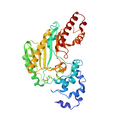
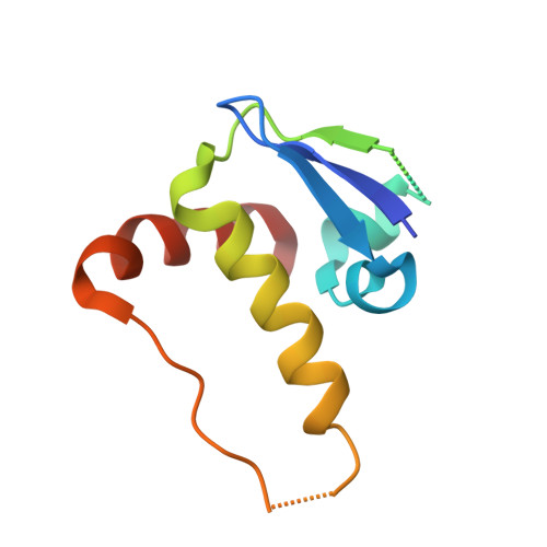
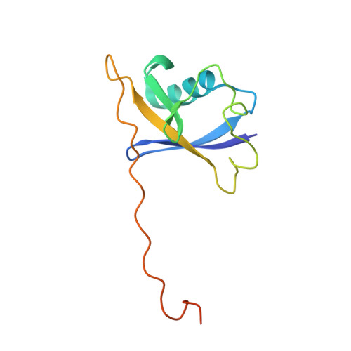
Structure and dynamics of the ASB9 CUL-RING E3 Ligase.

Lumpkin, R.J.Baker, R.W.Leschziner, A.E.Komives, E.A.

(2020) Nat Commun 11: 2866-2866

  • DOI: https://doi.org/10.1038/s41467-020-16499-9
  • Primary Citation Related Structures: 
    6V9H, 6V9I

  • PubMed Abstract: 

    The Cullin 5 (CUL5) Ring E3 ligase uses adaptors Elongins B and C (ELOB/C) to bind different SOCS-box-containing substrate receptors, determining the substrate specificity of the ligase. The 18-member ankyrin and SOCS box (ASB) family is the largest substrate receptor family. Here we report cryo-EM data for the substrate, creatine kinase (CKB) bound to ASB9-ELOB/C, and for full-length CUL5 bound to the RING protein, RBX2, which binds various E2s. To date, no full structures are available either for a substrate-bound ASB nor for CUL5. Hydrogen-deuterium exchange (HDX-MS) mapped onto a full structural model of the ligase revealed long-range allostery extending from the substrate through CUL5. We propose a revised allosteric mechanism for how CUL-E3 ligases function. ASB9 and CUL5 behave as rigid rods, connected through a hinge provided by ELOB/C transmitting long-range allosteric crosstalk from the substrate through CUL5 to the RBX2 flexible linker.


  • Organizational Affiliation
    • Department of Chemistry and Biochemistry, University of California, San Diego, 9500 Gilman Drive, La Jolla, CA, 92092-0378, USA.

Macromolecule Content 

  • Total Structure Weight: 143.71 kDa 
  • Atom Count: 9,326 
  • Modeled Residue Count: 1,185 
  • Deposited Residue Count: 1,291 
  • Unique protein chains: 4

Macromolecules

Find similar proteins by:|  3D Structure
Entity ID: 1
MoleculeChains  Sequence LengthOrganismDetailsImage
Creatine kinase B-type
A, B
381Homo sapiensMutation(s): 0 
Gene Names: CKBCKBB
EC: 2.7.3.2
UniProt & NIH Common Fund Data Resources
Find proteins for P12277 (Homo sapiens)
Explore P12277 
Go to UniProtKB:  P12277
PHAROS:  P12277
GTEx:  ENSG00000166165 
Entity Groups
Sequence Clusters30% Identity50% Identity70% Identity90% Identity95% Identity100% Identity
UniProt GroupP12277
Sequence Annotations
Expand
Reference Sequence
Find similar proteins by:|  3D Structure
Entity ID: 2
MoleculeChains  Sequence LengthOrganismDetailsImage
Ankyrin repeat and SOCS box protein 9314Homo sapiensMutation(s): 0 
Gene Names: ASB9
UniProt & NIH Common Fund Data Resources
Find proteins for Q96DX5 (Homo sapiens)
Explore Q96DX5 
Go to UniProtKB:  Q96DX5
PHAROS:  Q96DX5
GTEx:  ENSG00000102048 
Entity Groups
Sequence Clusters30% Identity50% Identity70% Identity90% Identity95% Identity100% Identity
UniProt GroupQ96DX5
Sequence Annotations
Expand
Reference Sequence
Find similar proteins by:|  3D Structure
Entity ID: 3
MoleculeChains  Sequence LengthOrganismDetailsImage
Elongin-C97Homo sapiensMutation(s): 0 
Gene Names: ELOCTCEB1
UniProt & NIH Common Fund Data Resources
Find proteins for Q15369 (Homo sapiens)
Explore Q15369 
Go to UniProtKB:  Q15369
PHAROS:  Q15369
GTEx:  ENSG00000154582 
Entity Groups
Sequence Clusters30% Identity50% Identity70% Identity90% Identity95% Identity100% Identity
UniProt GroupQ15369
Sequence Annotations
Expand
Reference Sequence
Find similar proteins by:|  3D Structure
Entity ID: 4
MoleculeChains  Sequence LengthOrganismDetailsImage
Elongin-B118Homo sapiensMutation(s): 0 
Gene Names: ELOBTCEB2
UniProt & NIH Common Fund Data Resources
Find proteins for Q15370 (Homo sapiens)
Explore Q15370 
Go to UniProtKB:  Q15370
PHAROS:  Q15370
GTEx:  ENSG00000103363 
Entity Groups
Sequence Clusters30% Identity50% Identity70% Identity90% Identity95% Identity100% Identity
UniProt GroupQ15370
Sequence Annotations
Expand
Reference Sequence

Experimental Data & Validation

Experimental Data

  • Method: ELECTRON MICROSCOPY
  • Resolution: 4.10 Å
  • Aggregation State: PARTICLE 
  • Reconstruction Method: SINGLE PARTICLE 
EM Software:
TaskSoftware PackageVersion
RECONSTRUCTIONcryoSPARC2
MODEL REFINEMENTRosetta

Structure Validation

View Full Validation Report


Entry History 

& Funding Information

Deposition Data


Funding OrganizationLocationGrant Number
National Science Foundation (NSF, United States)United States1817774

Revision History  (Full details and data files)

  • Version 1.0: 2020-04-29
    Type: Initial release
  • Version 1.1: 2020-11-11
    Changes: Database references
  • Version 1.2: 2024-10-30
    Changes: Data collection, Database references, Refinement description, Structure summary