6V6W | pdb_00006v6w

Crystal structure of antibody 438-B11 DSS mutant (Cys98A-100aA) in complex with an uncleaved prefusion optimized (UFO) soluble BG505 trimer and Fab 35O22


Experimental Data Snapshot

  • Method: X-RAY DIFFRACTION
  • Resolution: 6.50 Å
  • R-Value Free: 
    0.355 (Depositor), 0.355 (DCC) 
  • R-Value Work: 
    0.309 (Depositor), 0.310 (DCC) 
  • R-Value Observed: 
    0.311 (Depositor) 

Starting Model: experimental
View more details

wwPDB Validation 3D Report Full Report

Validation slider image for 6V6W

Ligand Structure Quality Assessment 


This is version 1.3 of the entry. See complete history

Literature

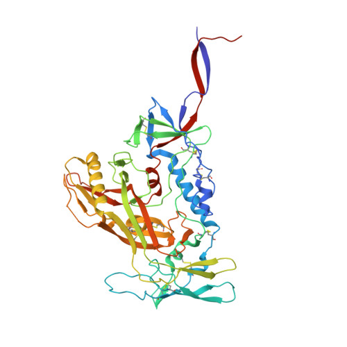
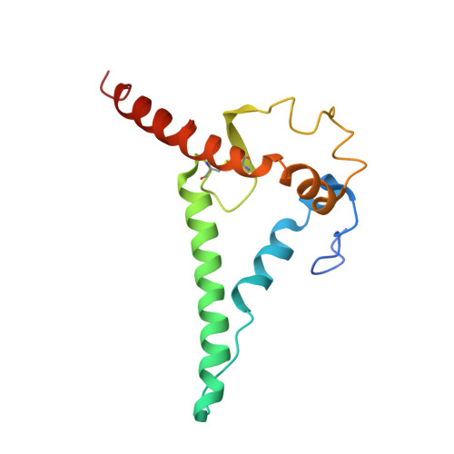
A VH1-69 antibody lineage from an infected Chinese donor potently neutralizes HIV-1 by targeting the V3 glycan supersite

Kumar, S.Ju, B.Shapero, B.Lin, X.Ren, L.Zhang, L.Li, D.Zhou, Z.Feng, Y.Sou, C.Mann, C.J.Hao, Y.Sarkar, A.Hou, J.Nunnally, C.Hong, K.Wang, S.Ge, X.Su, B.Landais, E.Sok, D.Zwick, M.B.He, L.Zhu, J.Wilson, I.A.Shao, Y.

(2020) Sci Adv 6: eabb1328

Macromolecule Content 

  • Total Structure Weight: 344.68 kDa 
  • Atom Count: 23,434 
  • Modeled Residue Count: 2,958 
  • Deposited Residue Count: 3,068 
  • Unique protein chains: 6

Macromolecules

Find similar proteins by:|  3D Structure
Entity ID: 1
MoleculeChains  Sequence LengthOrganismDetailsImage
35O22 Fab Heavy chainA [auth D],
G [auth A]
243Human immunodeficiency virus 1Mutation(s): 0 
Find similar proteins by:|  3D Structure
Entity ID: 2
MoleculeChains  Sequence LengthOrganismDetailsImage
35O22 Fab Light chainB [auth E],
H [auth B]
216Human immunodeficiency virus 1Mutation(s): 0 
Find similar proteins by:|  3D Structure
Entity ID: 3
MoleculeChains  Sequence LengthOrganismDetailsImage
Envelope glycoprotein gp120C [auth G],
I [auth C]
485Human immunodeficiency virus 1Mutation(s): 0 
UniProt
Find proteins for Q2N0S6 (Human immunodeficiency virus type 1)
Explore Q2N0S6 
Go to UniProtKB:  Q2N0S6
Entity Groups
Sequence Clusters30% Identity50% Identity70% Identity90% Identity95% Identity100% Identity
UniProt GroupQ2N0S6
Glycosylation
Glycosylation Sites: 12
Sequence Annotations
Expand
Reference Sequence
Find similar proteins by:|  3D Structure
Entity ID: 4
MoleculeChains  Sequence LengthOrganismDetailsImage
B11 DSS Fab heavy chainD [auth I],
J [auth F]
235Human immunodeficiency virus 1Mutation(s): 0 
Find similar proteins by:|  3D Structure
Entity ID: 5
MoleculeChains  Sequence LengthOrganismDetailsImage
B11 Fab light chainE [auth J],
K [auth H]
215Human immunodeficiency virus 1Mutation(s): 0 
Find similar proteins by:|  3D Structure
Entity ID: 6
MoleculeChains  Sequence LengthOrganismDetailsImage
Envelope glycoprotein gp41F [auth T],
L [auth K]
140Human immunodeficiency virus 1Mutation(s): 0 
Gene Names: env
UniProt
Find proteins for Q2N0S9 (Human immunodeficiency virus type 1)
Explore Q2N0S9 
Go to UniProtKB:  Q2N0S9
Entity Groups
Sequence Clusters30% Identity50% Identity70% Identity90% Identity95% Identity100% Identity
UniProt GroupQ2N0S9
Glycosylation
Glycosylation Sites: 3
Sequence Annotations
Expand
Reference Sequence

Oligosaccharides

Help  
Entity ID: 7
MoleculeChains Length2D Diagram GlycosylationD Interactions
alpha-D-mannopyranose-(1-6)-beta-D-mannopyranose-(1-4)-2-acetamido-2-deoxy-beta-D-glucopyranose-(1-4)-2-acetamido-2-deoxy-beta-D-glucopyranoseM [auth L],
P [auth O]
4N-Glycosylation
Glycosylation Resources
GlyTouCan: G22573RC
GlyCosmos: G22573RC
GlyGen: G22573RC
Entity ID: 8
MoleculeChains Length2D Diagram GlycosylationD Interactions
2-acetamido-2-deoxy-beta-D-glucopyranose-(1-4)-2-acetamido-2-deoxy-beta-D-glucopyranoseN [auth M],
Q [auth P]
2N-Glycosylation
Glycosylation Resources
GlyTouCan: G42666HT
GlyCosmos: G42666HT
GlyGen: G42666HT
Entity ID: 9
MoleculeChains Length2D Diagram GlycosylationD Interactions
alpha-D-mannopyranose-(1-3)-[alpha-D-mannopyranose-(1-6)]beta-D-mannopyranose-(1-4)-2-acetamido-2-deoxy-beta-D-glucopyranose-(1-4)-2-acetamido-2-deoxy-beta-D-glucopyranoseO [auth N],
R [auth Q]
5N-Glycosylation
Glycosylation Resources
GlyTouCan: G22768VO
GlyCosmos: G22768VO
GlyGen: G22768VO

Small Molecules

Ligands 1 Unique
IDChains Name / Formula / InChI Key2D Diagram3D Interactions
NAG

Query on NAG


Download:Ideal Coordinates CCD File
AA [auth G]
BA [auth T]
CA [auth T]
DA [auth T]
EA [auth C]
AA [auth G],
BA [auth T],
CA [auth T],
DA [auth T],
EA [auth C],
FA [auth C],
GA [auth C],
HA [auth C],
IA [auth C],
JA [auth C],
KA [auth C],
LA [auth C],
MA [auth C],
NA [auth K],
OA [auth K],
PA [auth K],
S [auth G],
T [auth G],
U [auth G],
V [auth G],
W [auth G],
X [auth G],
Y [auth G],
Z [auth G]
2-acetamido-2-deoxy-beta-D-glucopyranose
C8 H15 N O6
OVRNDRQMDRJTHS-FMDGEEDCSA-N

Experimental Data & Validation

Experimental Data

  • Method: X-RAY DIFFRACTION
  • Resolution: 6.50 Å
  • R-Value Free:  0.355 (Depositor), 0.355 (DCC) 
  • R-Value Work:  0.309 (Depositor), 0.310 (DCC) 
  • R-Value Observed: 0.311 (Depositor) 
Space Group: H 3
Unit Cell:
Length ( Å )Angle ( ˚ )
a = 148.534α = 90
b = 148.534β = 90
c = 843.554γ = 120
Software Package:
Software NamePurpose
PHENIXrefinement
Cootmodel building
HKL-2000data scaling
HKL-2000data processing
HKL-2000data reduction
PHASERphasing

Structure Validation

View Full Validation Report


Ligand Structure Quality Assessment 


Entry History 

Deposition Data

Revision History  (Full details and data files)

  • Version 1.0: 2020-09-23
    Type: Initial release
  • Version 1.1: 2021-01-27
    Changes: Derived calculations
  • Version 1.2: 2023-10-11
    Changes: Data collection, Database references, Refinement description
  • Version 1.3: 2024-10-23
    Changes: Structure summary