6UUI | pdb_00006uui

Crystal structure of the heterocomplex between coil 2B domains of wild-type keratin 1 (KRT1) and keratin 10 (KRT10) containing mutation Cys401Ala


Experimental Data Snapshot

  • Method: X-RAY DIFFRACTION
  • Resolution: 2.07 Å
  • R-Value Free: 
    0.277 (Depositor), 0.283 (DCC) 
  • R-Value Work: 
    0.250 (Depositor), 0.258 (DCC) 
  • R-Value Observed: 
    0.252 (Depositor) 

Starting Model: experimental
View more details

wwPDB Validation 3D Report Full Report

Validation slider image for 6UUI

Ligand Structure Quality Assessment 


This is version 1.4 of the entry. See complete history

Literature

Crystal Structure of Keratin 1/10(C401A) 2B Heterodimer Demonstrates a Proclivity for the C-Terminus of Helix 2B to Form Higher Order Molecular Contacts.

Lomakin, I.B.Hinbest, A.J.Ho, M.Eldirany, S.A.Bunick, C.G.

(2020) Yale J Biol Med 93: 3-17

  • Primary Citation Related Structures: 
    6UUI

  • PubMed Abstract: 

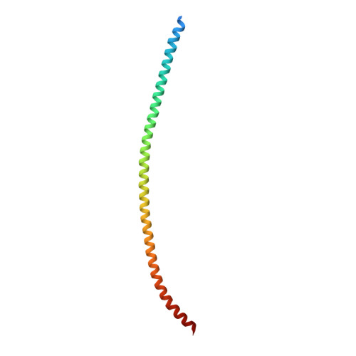
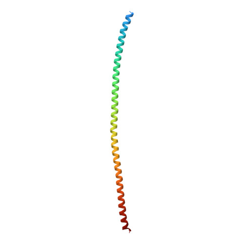
    We previously determined the crystal structure of the wild-type keratin 1/10 helix 2B heterodimer at 3.3 Å resolution. We proposed that the resolution of the diffraction data was limited due to the crystal packing effect from keratin 10 (K10) residue Cys401. Cys401 K10 formed a disulfide-linkage with Cys401 from another K1/10 heterodimer, creating an "X-shaped" structure and a loose crystal packing arrangement. We hypothesized that mutation of Cys401 K10 to alanine would eliminate the disulfide-linkage and improve crystal packing thereby increasing resolution of diffraction and enabling a more accurate side chain electron density map. Indeed, when a K10 Cys401Ala 2B mutant was paired with its native keratin 1 (K1) 2B heterodimer partner its x-ray crystal structure was determined at 2.07 Å resolution; the structure does not contain a disulfide linkage. Superposition of the K1/K10(Cys401Ala) 2B structure onto the wild-type K1/10 2B heterodimer structure had a root-mean-square-deviation of 1.88 Å; the variability in the atomic positions reflects the dynamic motion expected in this filamentous coiled-coil complex. The electrostatic, hydrophobic, and contour features of the molecular surface are similar to the lower resolution wild-type structure. We postulated that elimination of the disulfide linkage in the K1/K10(Cys401Ala) 2B structure could allow for the 2B heterodimers to bind/pack in the A 22 tetramer configuration associated with mature keratin intermediate filament assembly. Analysis of the crystal packing revealed a half-staggered anti-parallel tetrameric complex of 2B heterodimers; however, their register is not consistent with models of the A 22 mode of tetrameric alignment or prior biochemical cross-linking studies.


  • Organizational Affiliation
    • Department of Molecular Biophysics and Biochemistry, Yale University, New Haven, CT.

Macromolecule Content 

  • Total Structure Weight: 28.34 kDa 
  • Atom Count: 1,886 
  • Modeled Residue Count: 215 
  • Deposited Residue Count: 242 
  • Unique protein chains: 2

Macromolecules

Find similar proteins by:|  3D Structure
Entity ID: 1
MoleculeChains  Sequence LengthOrganismDetailsImage
Keratin, type II cytoskeletal 1A [auth C]120Homo sapiensMutation(s): 0 
Gene Names: KRT1KRTA
UniProt & NIH Common Fund Data Resources
Find proteins for P04264 (Homo sapiens)
Explore P04264 
Go to UniProtKB:  P04264
PHAROS:  P04264
GTEx:  ENSG00000167768 
Entity Groups
Sequence Clusters30% Identity50% Identity70% Identity90% Identity95% Identity100% Identity
UniProt GroupP04264
Sequence Annotations
Expand
Reference Sequence
Find similar proteins by:|  3D Structure
Entity ID: 2
MoleculeChains  Sequence LengthOrganismDetailsImage
Keratin, type I cytoskeletal 10B [auth X]122Homo sapiensMutation(s): 1 
Gene Names: KRT10KPP
UniProt & NIH Common Fund Data Resources
Find proteins for P13645 (Homo sapiens)
Explore P13645 
Go to UniProtKB:  P13645
PHAROS:  P13645
GTEx:  ENSG00000186395 
Entity Groups
Sequence Clusters30% Identity50% Identity70% Identity90% Identity95% Identity100% Identity
UniProt GroupP13645
Sequence Annotations
Expand
Reference Sequence

Small Molecules

Ligands 2 Unique
IDChains Name / Formula / InChI Key2D Diagram3D Interactions
BOG

Query on BOG


Download:Ideal Coordinates CCD File
C
octyl beta-D-glucopyranoside
C14 H28 O6
HEGSGKPQLMEBJL-RKQHYHRCSA-N
GOL

Query on GOL


Download:Ideal Coordinates CCD File
D [auth X]GLYCEROL
C3 H8 O3
PEDCQBHIVMGVHV-UHFFFAOYSA-N

Experimental Data & Validation

Experimental Data

  • Method: X-RAY DIFFRACTION
  • Resolution: 2.07 Å
  • R-Value Free:  0.277 (Depositor), 0.283 (DCC) 
  • R-Value Work:  0.250 (Depositor), 0.258 (DCC) 
  • R-Value Observed: 0.252 (Depositor) 
Space Group: P 61 2 2
Unit Cell:
Length ( Å )Angle ( ˚ )
a = 100.124α = 90
b = 100.124β = 90
c = 224.788γ = 120
Software Package:
Software NamePurpose
PHENIXrefinement
HKL-2000data reduction
HKL-2000data scaling
MOLREPphasing
PDB_EXTRACTdata extraction

Structure Validation

View Full Validation Report


Ligand Structure Quality Assessment 


Entry History 

& Funding Information

Deposition Data


Funding OrganizationLocationGrant Number
National Institutes of Health/National Institute of Arthritis and Musculoskeletal and Skin Diseases (NIH/NIAMS)United StatesK08AR070290
Other privateUnited StatesFoundation for Ichthyosis and Related Skin Types

Revision History  (Full details and data files)

  • Version 1.0: 2019-11-13
    Type: Initial release
  • Version 1.1: 2019-12-11
    Changes: Author supporting evidence, Database references
  • Version 1.2: 2020-04-08
    Changes: Database references
  • Version 1.3: 2020-07-29
    Type: Remediation
    Reason: Carbohydrate remediation
    Changes: Data collection, Derived calculations, Structure summary
  • Version 1.4: 2023-10-11
    Changes: Data collection, Database references, Refinement description, Structure summary