6UTB | pdb_00006utb

CRYSTAL STRUCTURE OF UNLIGANDED HIV-1 LM/HT CLADE A/E CRF01 GP120 CORE


Experimental Data Snapshot

  • Method: X-RAY DIFFRACTION
  • Resolution: 2.50 Å
  • R-Value Free: 
    0.305 (Depositor), 0.317 (DCC) 
  • R-Value Work: 
    0.263 (Depositor), 0.265 (DCC) 
  • R-Value Observed: 
    0.265 (Depositor) 

Starting Model: experimental
View more details

wwPDB Validation 3D Report Full Report

Validation slider image for 6UTB

Ligand Structure Quality Assessment 


This is version 1.2 of the entry. See complete history

Literature

The HIV-1 Env gp120 Inner Domain Shapes the Phe43 Cavity and the CD4 Binding Site.

Prevost, J.Tolbert, W.D.Medjahed, H.Sherburn, R.T.Madani, N.Zoubchenok, D.Gendron-Lepage, G.Gaffney, A.E.Grenier, M.C.Kirk, S.Vergara, N.Han, C.Mann, B.T.Chenine, A.L.Ahmed, A.Chaiken, I.Kirchhoff, F.Hahn, B.H.Haim, H.Abrams, C.F.Smith 3rd, A.B.Sodroski, J.Pazgier, M.Finzi, A.

(2020) mBio 11

  • DOI: https://doi.org/10.1128/mBio.00280-20
  • Primary Citation Related Structures: 
    6USW, 6UT1, 6UTB, 6UTD

  • PubMed Abstract: 

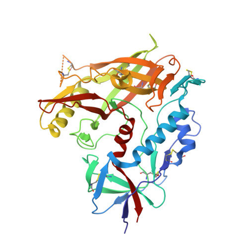
    The HIV-1 envelope glycoproteins (Env) undergo conformational changes upon interaction of the gp120 exterior glycoprotein with the CD4 receptor. The gp120 inner domain topological layers facilitate the transition of Env to the CD4-bound conformation. CD4 engages gp120 by introducing its phenylalanine 43 (Phe43) in a cavity ("the Phe43 cavity") located at the interface between the inner and outer gp120 domains. Small CD4-mimetic compounds (CD4mc) can bind within the Phe43 cavity and trigger conformational changes similar to those induced by CD4. Crystal structures of CD4mc in complex with a modified CRF01_AE gp120 core revealed the importance of these gp120 inner domain layers in stabilizing the Phe43 cavity and shaping the CD4 binding site. Our studies reveal a complex interplay between the gp120 inner domain and the Phe43 cavity and generate useful information for the development of more-potent CD4mc. IMPORTANCE The Phe43 cavity of HIV-1 envelope glycoproteins (Env) is an attractive druggable target. New promising compounds, including small CD4 mimetics (CD4mc), were shown to insert deeply into this cavity. Here, we identify a new network of residues that helps to shape this highly conserved CD4 binding pocket and characterize the structural determinants responsible for Env sensitivity to small CD4 mimetics.


  • Organizational Affiliation
    • Centre de Recherche du CHUM, Montreal, Quebec, Canada.

Macromolecule Content 

  • Total Structure Weight: 41.92 kDa 
  • Atom Count: 2,875 
  • Modeled Residue Count: 341 
  • Deposited Residue Count: 355 
  • Unique protein chains: 1

Macromolecules

Find similar proteins by:|  3D Structure
Entity ID: 1
MoleculeChains  Sequence LengthOrganismDetailsImage
HIV-1 LM/HT Clade A/E CRF01 gp120 core355Human immunodeficiency virus 1Mutation(s): 7 
Gene Names: HIV-1 Env
UniProt
Find proteins for A0A0M3KKW9 (Human immunodeficiency virus type 1)
Explore A0A0M3KKW9 
Go to UniProtKB:  A0A0M3KKW9
Entity Groups
Sequence Clusters30% Identity50% Identity70% Identity90% Identity95% Identity100% Identity
UniProt GroupA0A0M3KKW9
Glycosylation
Glycosylation Sites: 10
Sequence Annotations
Expand
Reference Sequence

Small Molecules

Ligands 2 Unique
IDChains Name / Formula / InChI Key2D Diagram3D Interactions
EPE

Query on EPE


Download:Ideal Coordinates CCD File
L [auth A]4-(2-HYDROXYETHYL)-1-PIPERAZINE ETHANESULFONIC ACID
C8 H18 N2 O4 S
JKMHFZQWWAIEOD-UHFFFAOYSA-N
NAG

Query on NAG


Download:Ideal Coordinates CCD File
B [auth A]
C [auth A]
D [auth A]
E [auth A]
F [auth A]
B [auth A],
C [auth A],
D [auth A],
E [auth A],
F [auth A],
G [auth A],
H [auth A],
I [auth A],
J [auth A],
K [auth A]
2-acetamido-2-deoxy-beta-D-glucopyranose
C8 H15 N O6
OVRNDRQMDRJTHS-FMDGEEDCSA-N

Experimental Data & Validation

Experimental Data

  • Method: X-RAY DIFFRACTION
  • Resolution: 2.50 Å
  • R-Value Free:  0.305 (Depositor), 0.317 (DCC) 
  • R-Value Work:  0.263 (Depositor), 0.265 (DCC) 
  • R-Value Observed: 0.265 (Depositor) 
Space Group: P 21 21 21
Unit Cell:
Length ( Å )Angle ( ˚ )
a = 66.901α = 90
b = 67.687β = 90
c = 86.47γ = 90
Software Package:
Software NamePurpose
PHENIXrefinement
XDSdata reduction
XDSdata scaling
PHASERphasing

Structure Validation

View Full Validation Report


Ligand Structure Quality Assessment 


Entry History 

& Funding Information

Deposition Data


Funding OrganizationLocationGrant Number
National Institutes of Health/National Institute Of Allergy and Infectious Diseases (NIH/NIAID)United StatesR01 AI116274
National Institutes of Health/National Institute Of Allergy and Infectious Diseases (NIH/NIAID)United StatesP01 AI120756
National Institutes of Health/National Institute Of Allergy and Infectious Diseases (NIH/NIAID)United StatesR01 AI129769

Revision History  (Full details and data files)

  • Version 1.0: 2020-10-07
    Type: Initial release
  • Version 1.1: 2023-10-11
    Changes: Data collection, Database references, Refinement description
  • Version 1.2: 2024-10-30
    Changes: Structure summary