Novel bacterial clade reveals origin of form I Rubisco.
Banda, D.M., Pereira, J.H., Liu, A.K., Orr, D.J., Hammel, M., He, C., Parry, M.A.J., Carmo-Silva, E., Adams, P.D., Banfield, J.F., Shih, P.M.(2020) Nat Plants 6: 1158-1166
- PubMed: 32868887 Search on PubMedSearch on PubMed Central
- DOI: https://doi.org/10.1038/s41477-020-00762-4
- Primary Citation Related Structures:
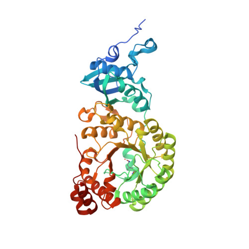
6URA - PubMed Abstract:
Rubisco sustains the biosphere through the fixation of CO 2 into biomass. In plants and cyanobacteria, form I Rubisco is structurally comprised of large and small subunits, whereas all other Rubisco forms lack small subunits. The rise of the form I complex through the innovation of small subunits represents a key, yet poorly understood, transition in Rubisco's evolution. Through metagenomic analyses, we discovered a previously uncharacterized clade sister to form I Rubisco that evolved without small subunits. This clade diverged before the evolution of cyanobacteria and the origin of the small subunit; thus, it provides a unique reference point to advance our understanding of form I Rubisco evolution. Structural and kinetic data presented here reveal how a proto-form I Rubisco assembled and functioned without the structural stability imparted from small subunits. Our findings provide insight into a key evolutionary transition of the most abundant enzyme on Earth and the predominant entry point for nearly all global organic carbon.
- Department of Plant Biology, University of California, Davis, Davis, CA, USA.
Organizational Affiliation: