6UR9 | pdb_00006ur9

DNA polymerase I Large Fragment from Bacillus stearothermophilus with DNA template, dideoxy primer, 3'-amino-ddGTP (nGTP), and Ca2+


Experimental Data Snapshot

  • Method: X-RAY DIFFRACTION
  • Resolution: 2.10 Å
  • R-Value Free: 
    0.243 (Depositor), 0.245 (DCC) 
  • R-Value Work: 
    0.204 (Depositor), 0.205 (DCC) 
  • R-Value Observed: 
    0.206 (Depositor) 

Starting Model: experimental
View more details

wwPDB Validation 3D Report Full Report

Validation slider image for 6UR9

Ligand Structure Quality Assessment 


This is version 1.2 of the entry. See complete history

Literature

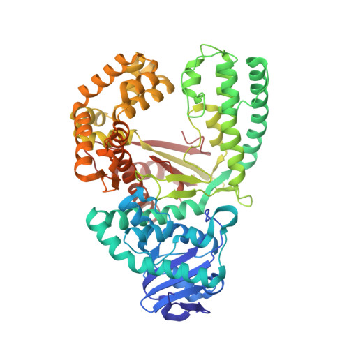
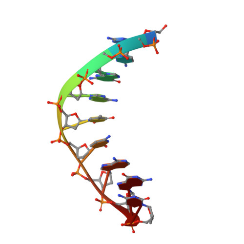
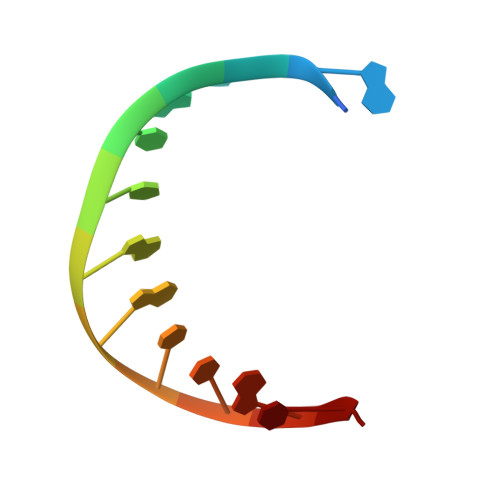
Synthesis of phosphoramidate-linked DNA by a modified DNA polymerase.

Lelyveld, V.S.Zhang, W.Szostak, J.W.

(2020) Proc Natl Acad Sci U S A 117: 7276-7283

  • DOI: https://doi.org/10.1073/pnas.1922400117
  • Primary Citation Related Structures: 
    6UR2, 6UR4, 6UR9, 6US5

  • PubMed Abstract: 

    All known polymerases copy genetic material by catalyzing phosphodiester bond formation. This highly conserved activity proceeds by a common mechanism, such that incorporated nucleoside analogs terminate chain elongation if the resulting primer strand lacks a terminal hydroxyl group. Even conservatively substituted 3'-amino nucleotides generally act as chain terminators, and no enzymatic pathway for their polymerization has yet been found. Although 3'-amino nucleotides can be chemically coupled to yield stable oligonucleotides containing N3'→P5' phosphoramidate (NP) bonds, no such internucleotide linkages are known to occur in nature. Here, we report that 3'-amino terminated primers are, in fact, slowly extended by the DNA polymerase from B. stearothermophilus in a template-directed manner. When its cofactor is Ca 2+ rather than Mg 2+ , the reaction is fivefold faster, permitting multiple turnover NP bond formation to yield NP-DNA strands from the corresponding 3'-amino-2',3'-dideoxynucleoside 5'-triphosphates. A single active site mutation further enhances the rate of NP-DNA synthesis by an additional 21-fold. We show that DNA-dependent NP-DNA polymerase activity depends on conserved active site residues and propose a likely mechanism for this activity based on a series of crystal structures of bound complexes. Our results significantly broaden the catalytic scope of polymerase activity and suggest the feasibility of a genetic transition between native nucleic acids and NP-DNA.


  • Organizational Affiliation
    • Department of Molecular Biology, Center for Computational and Integrative Biology, Massachusetts General Hospital, Boston, MA 02114.

Macromolecule Content 

  • Total Structure Weight: 146.88 kDa 
  • Atom Count: 10,551 
  • Modeled Residue Count: 1,202 
  • Deposited Residue Count: 1,202 
  • Unique protein chains: 1
  • Unique nucleic acid chains: 2

Macromolecules


Find similar proteins by:|  3D Structure
Entity ID: 1
MoleculeChains  Sequence LengthOrganismDetailsImage
DNA polymerase I
A, D
579Geobacillus stearothermophilusMutation(s): 2 
Gene Names: DPO1polA
EC: 2.7.7.7
UniProt
Find proteins for D9N168 (Geobacillus stearothermophilus)
Explore D9N168 
Go to UniProtKB:  D9N168
Entity Groups
Sequence Clusters30% Identity50% Identity70% Identity90% Identity95% Identity100% Identity
UniProt GroupD9N168
Sequence Annotations
Expand
Reference Sequence
Find similar nucleic acids by:  Sequence
Entity ID: 2
MoleculeChains LengthOrganismImage
DNA (5'-D(*GP*CP*GP*AP*TP*CP*AP*GP*(DOC))-3')
B, E
9synthetic construct
Sequence Annotations
Expand
Reference Sequence
Find similar nucleic acids by:  Sequence
Entity ID: 3
MoleculeChains LengthOrganismImage
DNA (5'-D(*CP*AP*CP*GP*CP*TP*GP*AP*TP*CP*GP*CP*A)-3')
C, F
13synthetic construct
Sequence Annotations
Expand
Reference Sequence

Small Molecules

Ligands 3 Unique
IDChains Name / Formula / InChI Key2D Diagram3D Interactions
NG3
(Subject of Investigation/LOI)

Query on NG3


Download:Ideal Coordinates CCD File
G [auth A]3'-amino-2',3'-dideoxyguanosine 5'-(tetrahydrogen triphosphate)
C10 H17 N6 O12 P3
ILTYANYMUGEMNF-KVQBGUIXSA-N
SO4

Query on SO4


Download:Ideal Coordinates CCD File
I [auth A]
J [auth A]
K [auth A]
L [auth A]
M [auth A]
I [auth A],
J [auth A],
K [auth A],
L [auth A],
M [auth A],
N [auth A],
O [auth A],
P [auth A],
Q [auth D]
SULFATE ION
O4 S
QAOWNCQODCNURD-UHFFFAOYSA-L
CA

Query on CA


Download:Ideal Coordinates CCD File
H [auth A]CALCIUM ION
Ca
BHPQYMZQTOCNFJ-UHFFFAOYSA-N

Experimental Data & Validation

Experimental Data

  • Method: X-RAY DIFFRACTION
  • Resolution: 2.10 Å
  • R-Value Free:  0.243 (Depositor), 0.245 (DCC) 
  • R-Value Work:  0.204 (Depositor), 0.205 (DCC) 
  • R-Value Observed: 0.206 (Depositor) 
Space Group: P 21 21 21
Unit Cell:
Length ( Å )Angle ( ˚ )
a = 93.629α = 90
b = 108.069β = 90
c = 149.748γ = 90
Software Package:
Software NamePurpose
PHENIXrefinement
PDB_EXTRACTdata extraction
HKL-2000data reduction
HKL-2000data scaling
PHASERphasing

Structure Validation

View Full Validation Report


Ligand Structure Quality Assessment 


Entry History 

& Funding Information

Deposition Data


Funding OrganizationLocationGrant Number
Howard Hughes Medical Institute (HHMI)United States--

Revision History  (Full details and data files)

  • Version 1.0: 2020-03-18
    Type: Initial release
  • Version 1.1: 2020-05-27
    Changes: Database references
  • Version 1.2: 2023-10-11
    Changes: Data collection, Database references, Derived calculations, Refinement description