6UQQ | pdb_00006uqq

Crystal Structure of GTPase Domain of Human Septin 7 / Septin 3 T282Y Heterocomplex


Experimental Data Snapshot

  • Method: X-RAY DIFFRACTION
  • Resolution: 2.75 Å
  • R-Value Free: 
    0.256 (Depositor), 0.253 (DCC) 
  • R-Value Work: 
    0.197 (Depositor), 0.197 (DCC) 
  • R-Value Observed: 
    0.200 (Depositor) 

Starting Models: experimental
View more details

wwPDB Validation 3D Report Full Report

Validation slider image for 6UQQ

Ligand Structure Quality Assessment 


This is version 1.4 of the entry. See complete history

Literature

Molecular Recognition at Septin Interfaces: The Switches Hold the Key.

Rosa, H.V.D.Leonardo, D.A.Brognara, G.Brandao-Neto, J.D'Muniz Pereira, H.Araujo, A.P.U.Garratt, R.C.

(2020) J Mol Biology 432: 5784-5801

  • DOI: https://doi.org/10.1016/j.jmb.2020.09.001
  • Primary Citation Related Structures: 
    6UPA, 6UPQ, 6UPR, 6UQQ

  • PubMed Abstract: 

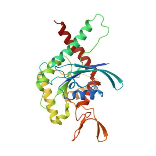
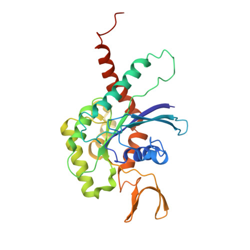
    The assembly of a septin filament requires that homologous monomers must distinguish between one another in establishing appropriate interfaces with their neighbors. To understand this phenomenon at the molecular level, we present the first four crystal structures of heterodimeric septin complexes. We describe in detail the two distinct types of G-interface present within the octameric particles, which must polymerize to form filaments. These are formed between SEPT2 and SEPT6 and between SEPT7 and SEPT3, and their description permits an understanding of the structural basis for the selectivity necessary for correct filament assembly. By replacing SEPT6 by SEPT8 or SEPT11, it is possible to rationalize Kinoshita's postulate, which predicts the exchangeability of septins from within a subgroup. Switches I and II, which in classical small GTPases provide a mechanism for nucleotide-dependent conformational change, have been repurposed in septins to play a fundamental role in molecular recognition. Specifically, it is switch I which holds the key to discriminating between the two different G-interfaces. Moreover, residues which are characteristic for a given subgroup play subtle, but pivotal, roles in guaranteeing that the correct interfaces are formed.


  • Organizational Affiliation
    • Instituto de Física de São Carlos, Universidade de São Paulo, Avenida João Dagnone 1100, São Carlos, SP 13563-723, Brazil.

Macromolecule Content 

  • Total Structure Weight: 135.12 kDa 
  • Atom Count: 8,453 
  • Modeled Residue Count: 1,049 
  • Deposited Residue Count: 1,156 
  • Unique protein chains: 2

Macromolecules

Find similar proteins by:|  3D Structure
Entity ID: 1
MoleculeChains  Sequence LengthOrganismDetailsImage
Septin-7
A, B
285Homo sapiensMutation(s): 0 
Gene Names: SEPT7CDC10
UniProt & NIH Common Fund Data Resources
Find proteins for Q16181 (Homo sapiens)
Explore Q16181 
Go to UniProtKB:  Q16181
PHAROS:  Q16181
GTEx:  ENSG00000122545 
Entity Groups
Sequence Clusters30% Identity50% Identity70% Identity90% Identity95% Identity100% Identity
UniProt GroupQ16181
Sequence Annotations
Expand
Reference Sequence
Find similar proteins by:|  3D Structure
Entity ID: 2
MoleculeChains  Sequence LengthOrganismDetailsImage
Neuronal-specific septin-3
C, D
293Homo sapiensMutation(s): 1 
Gene Names: SEPTIN3SEP3SEPT3
UniProt & NIH Common Fund Data Resources
Find proteins for Q9UH03 (Homo sapiens)
Explore Q9UH03 
Go to UniProtKB:  Q9UH03
PHAROS:  Q9UH03
GTEx:  ENSG00000100167 
Entity Groups
Sequence Clusters30% Identity50% Identity70% Identity90% Identity95% Identity100% Identity
UniProt GroupQ9UH03
Sequence Annotations
Expand
Reference Sequence

Experimental Data & Validation

Experimental Data

  • Method: X-RAY DIFFRACTION
  • Resolution: 2.75 Å
  • R-Value Free:  0.256 (Depositor), 0.253 (DCC) 
  • R-Value Work:  0.197 (Depositor), 0.197 (DCC) 
  • R-Value Observed: 0.200 (Depositor) 
Space Group: P 1 21 1
Unit Cell:
Length ( Å )Angle ( ˚ )
a = 61.39α = 90
b = 85.526β = 90.97
c = 152.051γ = 90
Software Package:
Software NamePurpose
Aimlessdata scaling
PHENIXrefinement
PDB_EXTRACTdata extraction
xia2data reduction
PHASERphasing

Structure Validation

View Full Validation Report


Ligand Structure Quality Assessment 


Entry History 

& Funding Information

Deposition Data


Funding OrganizationLocationGrant Number
Sao Paulo Research Foundation (FAPESP)Brazil2014/15546-1

Revision History  (Full details and data files)

  • Version 1.0: 2020-09-23
    Type: Initial release
  • Version 1.1: 2020-09-30
    Changes: Database references
  • Version 1.2: 2020-10-28
    Changes: Database references
  • Version 1.3: 2021-04-14
    Changes: Structure summary
  • Version 1.4: 2023-10-11
    Changes: Data collection, Database references, Refinement description