6UPW | pdb_00006upw

Metavinculin ABD-F-actin complex


Experimental Data Snapshot

  • Method: ELECTRON MICROSCOPY
  • Resolution: 2.90 Å
  • Aggregation State: HELICAL ARRAY 
  • Reconstruction Method: HELICAL 

Starting Models: experimental
View more details

wwPDB Validation 3D Report Full Report

Validation slider image for 6UPW

This is version 1.2 of the entry. See complete history

Literature

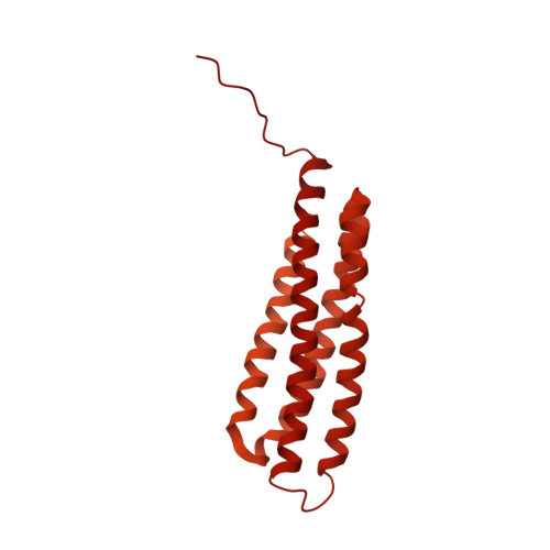
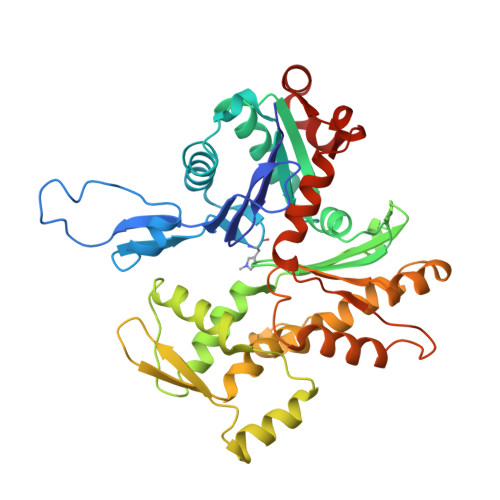
Molecular mechanism for direct actin force-sensing by alpha-catenin.

Mei, L.Espinosa de Los Reyes, S.Reynolds, M.J.Leicher, R.Liu, S.Alushin, G.M.

(2020) Elife 9

  • DOI: https://doi.org/10.7554/eLife.62514
  • Primary Citation Related Structures: 
    6UPV, 6UPW

  • PubMed Abstract: 

    The actin cytoskeleton mediates mechanical coupling between cells and their tissue microenvironments. The architecture and composition of actin networks are modulated by force; however, it is unclear how interactions between actin filaments (F-actin) and associated proteins are mechanically regulated. Here we employ both optical trapping and biochemical reconstitution with myosin motor proteins to show single piconewton forces applied solely to F-actin enhance binding by the human version of the essential cell-cell adhesion protein αE-catenin but not its homolog vinculin. Cryo-electron microscopy structures of both proteins bound to F-actin reveal unique rearrangements that facilitate their flexible C-termini refolding to engage distinct interfaces. Truncating α-catenin's C-terminus eliminates force-activated F-actin binding, and addition of this motif to vinculin confers force-activated binding, demonstrating that α-catenin's C-terminus is a modular detector of F-actin tension. Our studies establish that piconewton force on F-actin can enhance partner binding, which we propose mechanically regulates cellular adhesion through α-catenin.


  • Organizational Affiliation
    • Laboratory of Structural Biophysics and Mechanobiology, The Rockefeller University, New York, United States.

Macromolecule Content 

  • Total Structure Weight: 459.55 kDa 
  • Atom Count: 16,896 
  • Modeled Residue Count: 2,152 
  • Deposited Residue Count: 4,143 
  • Unique protein chains: 2

Macromolecules

Find similar proteins by:|  3D Structure
Entity ID: 1
MoleculeChains  Sequence LengthOrganismDetailsImage
VinculinA [auth L],
C [auth M]
1,134Homo sapiensMutation(s): 0 
Gene Names: VCL
UniProt & NIH Common Fund Data Resources
Find proteins for P18206 (Homo sapiens)
Explore P18206 
Go to UniProtKB:  P18206
PHAROS:  P18206
GTEx:  ENSG00000035403 
Entity Groups
Sequence Clusters30% Identity50% Identity70% Identity90% Identity95% Identity100% Identity
UniProt GroupP18206
Sequence Annotations
Expand
Reference Sequence
Find similar proteins by:|  3D Structure
Entity ID: 2
MoleculeChains  Sequence LengthOrganismDetailsImage
Actin, alpha skeletal muscleB [auth C],
D [auth B],
E [auth A],
F [auth D],
G [auth E]
375Gallus gallusMutation(s): 0 
EC: 3.6.4
UniProt
Find proteins for P68139 (Gallus gallus)
Explore P68139 
Go to UniProtKB:  P68139
Entity Groups
Sequence Clusters30% Identity50% Identity70% Identity90% Identity95% Identity100% Identity
UniProt GroupP68139
Sequence Annotations
Expand
Reference Sequence

Small Molecules

Ligands 2 Unique
IDChains Name / Formula / InChI Key2D Diagram3D Interactions
ADP

Query on ADP


Download:Ideal Coordinates CCD File
H [auth C],
J [auth B],
L [auth A],
N [auth D],
P [auth E]
ADENOSINE-5'-DIPHOSPHATE
C10 H15 N5 O10 P2
XTWYTFMLZFPYCI-KQYNXXCUSA-N
MG

Query on MG


Download:Ideal Coordinates CCD File
I [auth C],
K [auth B],
M [auth A],
O [auth D],
Q [auth E]
MAGNESIUM ION
Mg
JLVVSXFLKOJNIY-UHFFFAOYSA-N
Modified Residues  1 Unique
IDChains TypeFormula2D DiagramParent
HIC
Query on HIC
B [auth C],
D [auth B],
E [auth A],
F [auth D],
G [auth E]
L-PEPTIDE LINKINGC7 H11 N3 O2HIS

Experimental Data & Validation

Experimental Data

  • Method: ELECTRON MICROSCOPY
  • Resolution: 2.90 Å
  • Aggregation State: HELICAL ARRAY 
  • Reconstruction Method: HELICAL 
EM Software:
TaskSoftware PackageVersion
RECONSTRUCTIONRELION3.0
MODEL REFINEMENTPHENIX

Structure Validation

View Full Validation Report


Entry History 

& Funding Information

Deposition Data


Funding OrganizationLocationGrant Number
National Institutes of Health/National Institute of Dental and Craniofacial Research (NIH/NIDCR)United States5DP5OD017885

Revision History  (Full details and data files)

  • Version 1.0: 2020-09-30
    Type: Initial release
  • Version 1.1: 2020-10-07
    Changes: Database references
  • Version 1.2: 2025-04-02
    Changes: Data collection, Database references, Refinement description, Structure summary