6UPJ | pdb_00006upj

HIV-2 PROTEASE/U99294 COMPLEX


Experimental Data Snapshot

  • Method: X-RAY DIFFRACTION
  • Resolution: 2.34 Å
  • R-Value Work: 
    0.161 (Depositor) 

Starting Model: experimental
View more details

wwPDB Validation 3D Report Full Report

Validation slider image for 6UPJ

This is version 1.5 of the entry. See complete history

Literature

Use of medium-sized cycloalkyl rings to enhance secondary binding: discovery of a new class of human immunodeficiency virus (HIV) protease inhibitors.

Romines, K.R.Watenpaugh, K.D.Tomich, P.K.Howe, W.J.Morris, J.K.Lovasz, K.D.Mulichak, A.M.Finzel, B.C.Lynn, J.C.Horng, M.-M.Schwende, F.J.Ruwart, M.J.Zipp, G.L.Chong, K.-T.Dolak, L.A.Toth, L.N.Howard, G.M.Rush, B.D.Wilkinson, K.F.Possert, P.L.Dalga, R.J.Hinshaw, R.R.

(1995) J Med Chem 38: 1884-1891

  • DOI: https://doi.org/10.1021/jm00011a008
  • Primary Citation Related Structures: 
    5UPJ, 6UPJ

  • PubMed Abstract: 

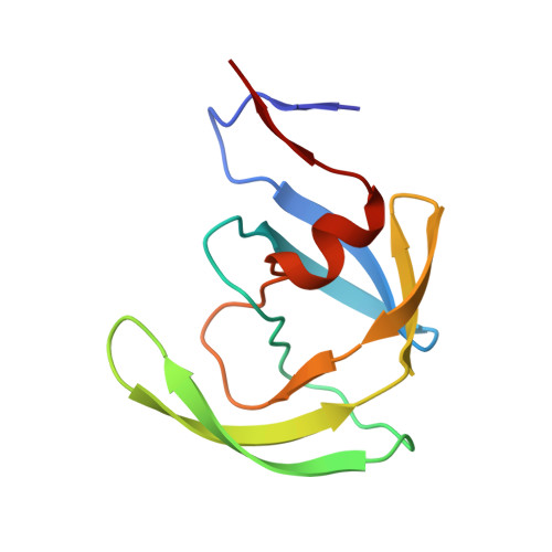
    A unique strategy for the enhancement of secondary binding of an inhibitor to an enzyme has been demonstrated in the design of new human immunodeficiency virus (HIV) protease inhibitors. When the planar benzene ring of a 4-hydroxycoumarin lead compound (1a, Ki = 0.800 microM) was replaced with medium-sized (i.e., 7-9), conformationally-flexible, alkyl rings, the enzyme inhibitory activity of the resulting compounds was dramatically improved, and inhibitors with more than 50-fold better binding (e.g., 5d, Ki = 0.015 microM) were obtained. X-ray crystal structures of these inhibitors complexed with HIV protease indicated the cycloalkyl rings were able to fold into the S1' pocket of the enzyme and fill it much more effectively than the rigid benzene ring of the 4-hydroxycoumarin compound. This work has resulted in the identification of a promising lead structure for the design of potent, deliverable HIV protease inhibitors. Compound 5d, a small (MW = 324), nonpeptidic structure, has already shown several advantages over peptidic inhibitors, including high oral bioavailability (91-99%), a relatively long half-life (4.9 h), and ease of synthesis (three steps).


  • Organizational Affiliation
    • Upjohn Laboratories, Kalamazoo, Michigan 49001, USA.

Macromolecule Content 

  • Total Structure Weight: 21.72 kDa 
  • Atom Count: 1,782 
  • Modeled Residue Count: 198 
  • Deposited Residue Count: 198 
  • Unique protein chains: 1

Macromolecules

Find similar proteins by:|  3D Structure
Entity ID: 1
MoleculeChains  Sequence LengthOrganismDetailsImage
HIV-2 PROTEASE
A, B
99Human immunodeficiency virus 2Mutation(s): 1 
EC: 3.4.23.16
UniProt
Find proteins for P04584 (Human immunodeficiency virus type 2 subtype A (isolate ROD))
Explore P04584 
Go to UniProtKB:  P04584
Entity Groups
Sequence Clusters30% Identity50% Identity70% Identity90% Identity95% Identity100% Identity
UniProt GroupP04584
Sequence Annotations
Expand
Reference Sequence

Small Molecules

Ligands 1 Unique
IDChains Name / Formula / InChI Key2D Diagram3D Interactions
NIU

Query on NIU


Download:Ideal Coordinates CCD File
C [auth A]6,7,8,9-TETRAHYDRO-4-HYDROXY-3-(1-PHENYLPROPYL)CYCLOHEPTA[B]PYRAN-2-ONE
C19 H22 O3
YKJXQZGJGDTEOC-AWEZNQCLSA-N

Experimental Data & Validation

Experimental Data

  • Method: X-RAY DIFFRACTION
  • Resolution: 2.34 Å
  • R-Value Work:  0.161 (Depositor) 
Space Group: P 21 21 21
Unit Cell:
Length ( Å )Angle ( ˚ )
a = 33.889α = 90
b = 45.675β = 90
c = 134.574γ = 90
Software Package:
Software NamePurpose
MERLOTphasing
CEDARrefinement
XENGENdata reduction
XENGENdata scaling

Structure Validation

View Full Validation Report


Entry History 

Deposition Data

Revision History  (Full details and data files)

  • Version 1.0: 1997-04-21
    Type: Initial release
  • Version 1.1: 2008-03-25
    Changes: Version format compliance
  • Version 1.2: 2011-07-13
    Changes: Version format compliance
  • Version 1.3: 2012-02-22
    Changes: Database references
  • Version 1.4: 2021-11-03
    Changes: Database references, Derived calculations
  • Version 1.5: 2024-04-03
    Changes: Data collection, Refinement description