6UNI | pdb_00006uni

Human CYP3A4 bound to an inhibitor


Experimental Data Snapshot

  • Method: X-RAY DIFFRACTION
  • Resolution: 2.60 Å
  • R-Value Free: 
    0.265 (Depositor), 0.266 (DCC) 
  • R-Value Work: 
    0.211 (Depositor), 0.218 (DCC) 
  • R-Value Observed: 
    0.217 (Depositor) 

Starting Model: experimental
View more details

wwPDB Validation 3D Report Full Report

Validation slider image for 6UNI

Ligand Structure Quality Assessment 


This is version 1.3 of the entry. See complete history

Literature

An increase in side-group hydrophobicity largely improves the potency of ritonavir-like inhibitors of CYP3A4.

Samuels, E.R.Sevrioukova, I.F.

(2020) Bioorg Med Chem 28: 115349-115349

  • DOI: https://doi.org/10.1016/j.bmc.2020.115349
  • Primary Citation Related Structures: 
    6UNE, 6UNG, 6UNH, 6UNI, 6UNJ, 6UNK, 6UNL, 6UNM

  • PubMed Abstract: 

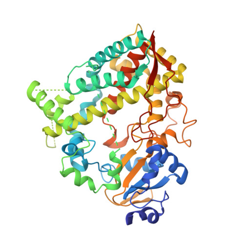
    Identification of structural determinants required for potent inhibition of drug-metabolizing cytochrome P450 3A4 (CYP3A4) could help develop safer drugs and more effective pharmacoenhancers. We utilize a rational inhibitor design to decipher structure-activity relationships in analogues of ritonavir, a highly potent CYP3A4 inhibitor marketed as pharmacoenhancer. Analysis of compounds with the R 1 side-group as phenyl or naphthalene and R 2 as indole or naphthalene in different stereo configuration showed that (i) analogues with the R 2 -naphthalene tend to bind tighter and inhibit CYP3A4 more potently than the R 2 -phenyl/indole containing counterparts; (ii) stereochemistry becomes a more important contributing factor, as the bulky side-groups limit the ability to optimize protein-ligand interactions; (iii) the relationship between the R 1 /R 2 configuration and preferential binding to CYP3A4 is complex and depends on the side-group functionality/interplay and backbone spacing; and (iv) three inhibitors, 5a-b and 7d, were superior to ritonavir (IC 50 of 0.055-0.085 μM vs. 0.130 μM, respectively).


  • Organizational Affiliation
    • Departments of Pharmaceutical Sciences, University of California, Irvine, CA 92697-3900, United States.

Macromolecule Content 

  • Total Structure Weight: 56.93 kDa 
  • Atom Count: 3,590 
  • Modeled Residue Count: 435 
  • Deposited Residue Count: 487 
  • Unique protein chains: 1

Macromolecules

Find similar proteins by:|  3D Structure
Entity ID: 1
MoleculeChains  Sequence LengthOrganismDetailsImage
Cytochrome P450 3A4487Homo sapiensMutation(s): 0 
Gene Names: CYP3A4CYP3A3
EC: 1.14.14.1 (PDB Primary Data), 1.14.14.56 (PDB Primary Data), 1.14.14.73 (PDB Primary Data), 1.14.14.55 (PDB Primary Data)
Membrane Entity: Yes 
UniProt & NIH Common Fund Data Resources
Find proteins for P08684 (Homo sapiens)
Explore P08684 
Go to UniProtKB:  P08684
PHAROS:  P08684
GTEx:  ENSG00000160868 
Entity Groups
Sequence Clusters30% Identity50% Identity70% Identity90% Identity95% Identity100% Identity
UniProt GroupP08684
Sequence Annotations
Expand
Reference Sequence

Small Molecules

Ligands 2 Unique
IDChains Name / Formula / InChI Key2D Diagram3D Interactions
HEM

Query on HEM


Download:Ideal Coordinates CCD File
B [auth A]PROTOPORPHYRIN IX CONTAINING FE
C34 H32 Fe N4 O4
KABFMIBPWCXCRK-RGGAHWMASA-L
QDV
(Subject of Investigation/LOI)

Query on QDV


Download:Ideal Coordinates CCD File
C [auth A]tert-butyl [(2S)-1-(1H-indol-3-yl)-3-{[(2R)-1-oxo-3-phenyl-1-{[2-(pyridin-3-yl)ethyl]amino}propan-2-yl]sulfanyl}propan-2-yl]carbamate
C32 H38 N4 O3 S
DCMGBSFQWSUWCW-LITSAYRRSA-N
Binding Affinity Annotations 
IDSourceBinding Affinity
QDV BindingDB:  6UNI IC50: 102 (nM) from 1 assay(s)

Experimental Data & Validation

Experimental Data

  • Method: X-RAY DIFFRACTION
  • Resolution: 2.60 Å
  • R-Value Free:  0.265 (Depositor), 0.266 (DCC) 
  • R-Value Work:  0.211 (Depositor), 0.218 (DCC) 
  • R-Value Observed: 0.217 (Depositor) 
Space Group: I 2 2 2
Unit Cell:
Length ( Å )Angle ( ˚ )
a = 76.036α = 90
b = 100.748β = 90
c = 125.229γ = 90
Software Package:
Software NamePurpose
SCALAdata scaling
PHENIXrefinement
PDB_EXTRACTdata extraction
XDSdata reduction
PHASERphasing

Structure Validation

View Full Validation Report


Ligand Structure Quality Assessment 


Entry History 

& Funding Information

Deposition Data


Funding OrganizationLocationGrant Number
National Institutes of Health/National Institute of Environmental Health Sciences (NIH/NIEHS)United StatesES025767

Revision History  (Full details and data files)

  • Version 1.0: 2020-02-05
    Type: Initial release
  • Version 1.1: 2020-02-26
    Changes: Database references
  • Version 1.2: 2020-03-04
    Changes: Database references
  • Version 1.3: 2023-10-11
    Changes: Data collection, Database references, Refinement description