6ULX | pdb_00006ulx

Adenylation domain of a keto acid-selecting NRPS module bound to keto acyl adenylate space group P43212


Experimental Data Snapshot

  • Method: X-RAY DIFFRACTION
  • Resolution: 2.31 Å
  • R-Value Free: 
    0.249 (Depositor), 0.254 (DCC) 
  • R-Value Work: 
    0.222 (Depositor), 0.227 (DCC) 
  • R-Value Observed: 
    0.223 (Depositor) 

wwPDB Validation 3D Report Full Report

Validation slider image for 6ULX

Ligand Structure Quality Assessment 


This is version 1.3 of the entry. See complete history

Literature

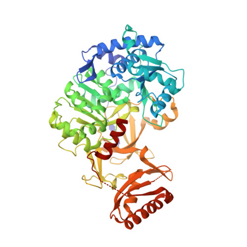
Structural basis of keto acid utilization in nonribosomal depsipeptide synthesis.

Alonzo, D.A.Chiche-Lapierre, C.Tarry, M.J.Wang, J.Schmeing, T.M.

(2020) Nat Chem Biol 16: 493-496

  • DOI: https://doi.org/10.1038/s41589-020-0481-5
  • Primary Citation Related Structures: 
    6ULW, 6ULX, 6ULY, 6ULZ

  • PubMed Abstract: 

    Nonribosomal depsipeptides are natural products composed of amino and hydroxy acid residues. The hydroxy acid residues often derive from α-keto acids, reduced by ketoreductase domains in the depsipeptide synthetases. Biochemistry and structures reveal the mechanism of discrimination for α-keto acids and a remarkable architecture: flanking intact adenylation and ketoreductase domains are sequences separated by >1,100 residues that form a split 'pseudoA sub ' domain, structurally important for the depsipeptide module's synthetic cycle.


  • Organizational Affiliation
    • Department of Biochemistry and Centre de Recherche en Biologie Structurale, McGill University, Montreal, Quebec, Canada.

Macromolecule Content 

  • Total Structure Weight: 66.03 kDa 
  • Atom Count: 4,470 
  • Modeled Residue Count: 551 
  • Deposited Residue Count: 576 
  • Unique protein chains: 1

Macromolecules

Find similar proteins by:|  3D Structure
Entity ID: 1
MoleculeChains  Sequence LengthOrganismDetailsImage
Amino acid adenylation domain-containing protein576Bacillus stratosphericus LAMA 585Mutation(s): 0 
Gene Names: C883_1060

Small Molecules

Ligands 2 Unique
IDChains Name / Formula / InChI Key2D Diagram3D Interactions
QA7
(Subject of Investigation/LOI)

Query on QA7


Download:Ideal Coordinates CCD File
B [auth A]5'-O-{(S)-hydroxy[(4-methyl-2-oxopentanoyl)oxy]phosphoryl}adenosine
C16 H22 N5 O9 P
QRKXCZFTEMACEI-SDBHATRESA-N
SO4

Query on SO4


Download:Ideal Coordinates CCD File
C [auth A]
D [auth A]
E [auth A]
F [auth A]
G [auth A]
C [auth A],
D [auth A],
E [auth A],
F [auth A],
G [auth A],
H [auth A],
I [auth A],
J [auth A],
K [auth A],
L [auth A],
M [auth A],
N [auth A],
O [auth A],
P [auth A],
Q [auth A],
R [auth A],
S [auth A]
SULFATE ION
O4 S
QAOWNCQODCNURD-UHFFFAOYSA-L

Experimental Data & Validation

Experimental Data

  • Method: X-RAY DIFFRACTION
  • Resolution: 2.31 Å
  • R-Value Free:  0.249 (Depositor), 0.254 (DCC) 
  • R-Value Work:  0.222 (Depositor), 0.227 (DCC) 
  • R-Value Observed: 0.223 (Depositor) 
Space Group: P 43 21 2
Unit Cell:
Length ( Å )Angle ( ˚ )
a = 103.274α = 90
b = 103.274β = 90
c = 172.747γ = 90
Software Package:
Software NamePurpose
PHENIXrefinement
DIALSdata reduction
Aimlessdata scaling
PHASERphasing

Structure Validation

View Full Validation Report


Ligand Structure Quality Assessment 


Entry History 

& Funding Information

Deposition Data


Funding OrganizationLocationGrant Number
Natural Sciences and Engineering Research Council (NSERC, Canada)Canada--

Revision History  (Full details and data files)

  • Version 1.0: 2020-02-19
    Type: Initial release
  • Version 1.1: 2020-03-04
    Changes: Database references
  • Version 1.2: 2020-05-06
    Changes: Database references
  • Version 1.3: 2024-03-13
    Changes: Data collection, Database references