6ULH | pdb_00006ulh

Structure of MavC in complex with its substrate in R3 spacegroup


Experimental Data Snapshot

  • Method: X-RAY DIFFRACTION
  • Resolution: 1.97 Å
  • R-Value Free: 
    0.227 (Depositor), 0.225 (DCC) 
  • R-Value Work: 
    0.183 (Depositor), 0.184 (DCC) 
  • R-Value Observed: 
    0.185 (Depositor) 

Starting Models: experimental
View more details

wwPDB Validation 3D Report Full Report

Validation slider image for 6ULH

This is version 1.2 of the entry. See complete history

Literature

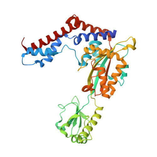
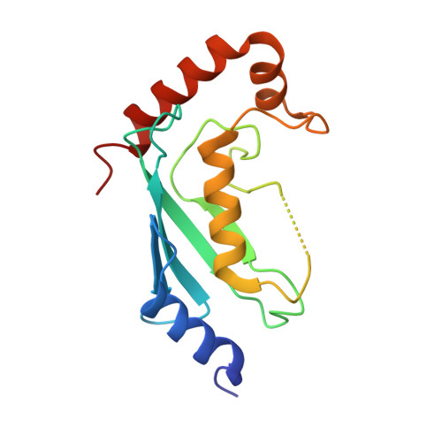
Legionella effector MavC targets the Ube2N~Ub conjugate for noncanonical ubiquitination.

Puvar, K.Iyer, S.Fu, J.Kenny, S.Negron Teron, K.I.Luo, Z.Q.Brzovic, P.S.Klevit, R.E.Das, C.

(2020) Nat Commun 11: 2365-2365

  • DOI: https://doi.org/10.1038/s41467-020-16211-x
  • Primary Citation Related Structures: 
    6P5B, 6P5H, 6ULH, 6UMP, 6UMS

  • PubMed Abstract: 

    The bacterial effector MavC modulates the host immune response by blocking Ube2N activity employing an E1-independent ubiquitin ligation, catalyzing formation of a γ-glutamyl-ε-Lys (Gln40 Ub -Lys92 Ube2N ) isopeptide crosslink using a transglutaminase mechanism. Here we provide biochemical evidence in support of MavC targeting the activated, thioester-linked Ube2N~ubiquitin conjugate, catalyzing an intramolecular transglutamination reaction, covalently crosslinking the Ube2N and Ub subunits effectively inactivating the E2~Ub conjugate. Ubiquitin exhibits weak binding to MavC alone, but shows an increase in affinity when tethered to Ube2N in a disulfide-linked substrate that mimics the charged E2~Ub conjugate. Crystal structures of MavC in complex with the substrate mimic and crosslinked product provide insights into the reaction mechanism and underlying protein dynamics that favor transamidation over deamidation, while revealing a crucial role for the structurally unique insertion domain in substrate recognition. This work provides a structural basis of ubiquitination by transglutamination and identifies this enzyme's true physiological substrate.


  • Organizational Affiliation
    • Department of Chemistry, Purdue University, West Lafayette, IN, 47907, USA.

Macromolecule Content 

  • Total Structure Weight: 69.98 kDa 
  • Atom Count: 4,906 
  • Modeled Residue Count: 597 
  • Deposited Residue Count: 617 
  • Unique protein chains: 3

Macromolecules

Find similar proteins by:|  3D Structure
Entity ID: 1
MoleculeChains  Sequence LengthOrganismDetailsImage
LPG2147 (MavC)389Legionella pneumophilaMutation(s): 1 
Gene Names: lpg2147
UniProt
Find proteins for Q5ZTL4 (Legionella pneumophila subsp. pneumophila (strain Philadelphia 1 / ATCC 33152 / DSM 7513))
Explore Q5ZTL4 
Go to UniProtKB:  Q5ZTL4
Entity Groups
Sequence Clusters30% Identity50% Identity70% Identity90% Identity95% Identity100% Identity
UniProt GroupQ5ZTL4
Sequence Annotations
Expand
Reference Sequence
Find similar proteins by:|  3D Structure
Entity ID: 2
MoleculeChains  Sequence LengthOrganismDetailsImage
Ubiquitin-conjugating enzyme E2 NB [auth C]152Homo sapiensMutation(s): 0 
Gene Names: UBE2NBLU
EC: 2.3.2.23
UniProt & NIH Common Fund Data Resources
Find proteins for P61088 (Homo sapiens)
Explore P61088 
Go to UniProtKB:  P61088
PHAROS:  P61088
GTEx:  ENSG00000177889 
Entity Groups
Sequence Clusters30% Identity50% Identity70% Identity90% Identity95% Identity100% Identity
UniProt GroupP61088
Sequence Annotations
Expand
Reference Sequence
Find similar proteins by:|  3D Structure
Entity ID: 3
MoleculeChains  Sequence LengthOrganismDetailsImage
UbiquitinC [auth E]76Homo sapiensMutation(s): 1 
Gene Names: UBC
UniProt & NIH Common Fund Data Resources
Find proteins for P0CG48 (Homo sapiens)
Explore P0CG48 
Go to UniProtKB:  P0CG48
PHAROS:  P0CG48
GTEx:  ENSG00000150991 
Entity Groups
Sequence Clusters30% Identity50% Identity70% Identity90% Identity95% Identity100% Identity
UniProt GroupP0CG48
Sequence Annotations
Expand
Reference Sequence

Experimental Data & Validation

Experimental Data

  • Method: X-RAY DIFFRACTION
  • Resolution: 1.97 Å
  • R-Value Free:  0.227 (Depositor), 0.225 (DCC) 
  • R-Value Work:  0.183 (Depositor), 0.184 (DCC) 
  • R-Value Observed: 0.185 (Depositor) 
Space Group: H 3
Unit Cell:
Length ( Å )Angle ( ˚ )
a = 171.859α = 90
b = 171.859β = 90
c = 58.362γ = 120
Software Package:
Software NamePurpose
PHENIXrefinement
TRUNCATEdata reduction
HKL-2000data scaling
PHENIXphasing

Structure Validation

View Full Validation Report


Entry History 

& Funding Information

Deposition Data


Funding OrganizationLocationGrant Number
National Institutes of Health/National Human Genome Research Institute (NIH/NHGRI)United States1R01GM126296

Revision History  (Full details and data files)

  • Version 1.0: 2020-05-27
    Type: Initial release
  • Version 1.1: 2023-10-11
    Changes: Advisory, Data collection, Database references, Refinement description
  • Version 1.2: 2024-10-16
    Changes: Structure summary