6UKH

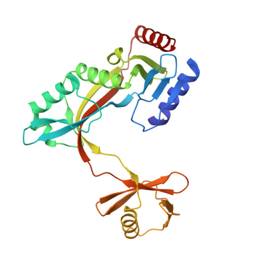
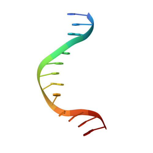
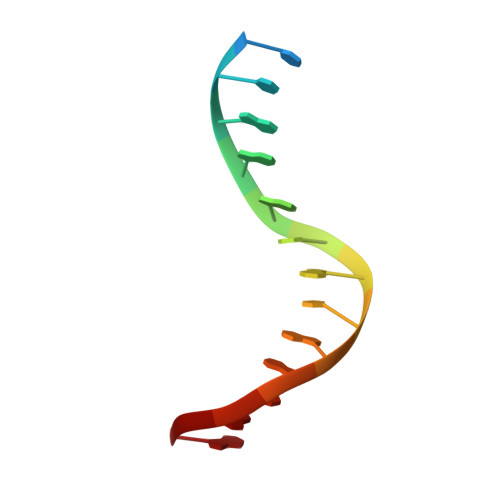
HhaI endonuclease in Complex with DNA in space group P41212 (pH 6.0)


Experimental Data Snapshot

  • Method: X-RAY DIFFRACTION
  • Resolution: 2.82 Å
  • R-Value Free: 0.252 
  • R-Value Work: 0.205 
  • R-Value Observed: 0.207 

Starting Model: experimental
View more details

wwPDB Validation   3D Report Full Report


This is version 1.3 of the entry. See complete history


Literature

Structure of HhaI endonuclease with cognate DNA at an atomic resolution of 1.0 angstrom.

Horton, J.R.Yang, J.Zhang, X.Petronzio, T.Fomenkov, A.Wilson, G.G.Roberts, R.J.Cheng, X.

(2020) Nucleic Acids Res 48: 1466-1478

  • DOI: https://doi.org/10.1093/nar/gkz1195
  • Primary Citation of Related Structures:  
    6UKE, 6UKF, 6UKG, 6UKH, 6UKI

  • PubMed Abstract: 

    HhaI, a Type II restriction endonuclease, recognizes the symmetric sequence 5'-GCG↓C-3' in duplex DNA and cleaves ('↓') to produce fragments with 2-base, 3'-overhangs. We determined the structure of HhaI in complex with cognate DNA at an ultra-high atomic resolution of 1.0 Å. Most restriction enzymes act as dimers with two catalytic sites, and cleave the two strands of duplex DNA simultaneously, in a single binding event. HhaI, in contrast, acts as a monomer with only one catalytic site, and cleaves the DNA strands sequentially, one after the other. HhaI comprises three domains, each consisting of a mixed five-stranded β sheet with a defined function. The first domain contains the catalytic-site; the second contains residues for sequence recognition; and the third contributes to non-specific DNA binding. The active-site belongs to the 'PD-D/EXK' superfamily of nucleases and contains the motif SD-X11-EAK. The first two domains are similar in structure to two other monomeric restriction enzymes, HinP1I (G↓CGC) and MspI (C↓CGG), which produce fragments with 5'-overhangs. The third domain, present only in HhaI, shifts the positions of the recognition residues relative to the catalytic site enabling this enzyme to cleave the recognition sequence at a different position. The structure of M.HhaI, the biological methyltransferase partner of HhaI, was determined earlier. Together, these two structures represent the first natural pair of restriction-modification enzymes to be characterized in atomic detail.


  • Organizational Affiliation

    Department of Epigenetics & Molecular Carcinogenesis, The University of Texas MD Anderson Cancer Center, Houston, TX 77030, USA.


Macromolecules

Find similar proteins by:  (by identity cutoff)  |  3D Structure
Entity ID: 1
MoleculeChains Sequence LengthOrganismDetailsImage
HhaI Restriction EndonucleaseA [auth X]258Haemophilus parahaemolyticusMutation(s): 0 
Gene Names: HMPREF1050_0931
EC: 3
Entity Groups  
Sequence Clusters30% Identity50% Identity70% Identity90% Identity95% Identity100% Identity
Sequence Annotations
Expand
  • Reference Sequence

Find similar nucleic acids by:  Sequence   |   3D Structure  

Entity ID: 2
MoleculeChains LengthOrganismImage
DNA (5'-D(*CP*TP*GP*TP*TP*GP*CP*GP*CP*TP*TP*GP*G)-3')B [auth A]14Haemophilus parahaemolyticus
Sequence Annotations
Expand
  • Reference Sequence

Find similar nucleic acids by:  Sequence   |   3D Structure  

Entity ID: 3
MoleculeChains LengthOrganismImage
DNA (5'-D(*CP*CP*AP*AP*GP*CP*GP*CP*AP*AP*CP*AP*G)-3')C [auth B]14Haemophilus parahaemolyticus
Sequence Annotations
Expand
  • Reference Sequence
Experimental Data & Validation

Experimental Data

  • Method: X-RAY DIFFRACTION
  • Resolution: 2.82 Å
  • R-Value Free: 0.252 
  • R-Value Work: 0.205 
  • R-Value Observed: 0.207 
  • Space Group: P 41 21 2
Unit Cell:
Length ( Å )Angle ( ˚ )
a = 67.257α = 90
b = 67.257β = 90
c = 171.072γ = 90
Software Package:
Software NamePurpose
SERGUIdata collection
PHENIXrefinement
HKL-2000data reduction
HKL-2000data scaling
PHENIXphasing

Structure Validation

View Full Validation Report


Entry History & Funding Information

Deposition Data


Funding OrganizationLocationGrant Number
National Institutes of Health/National Institute of General Medical Sciences (NIH/NIGMS)United StatesGM049245-23

Revision History  (Full details and data files)

  • Version 1.0: 2019-12-18
    Type: Initial release
  • Version 1.1: 2020-01-08
    Changes: Database references
  • Version 1.2: 2020-03-04
    Changes: Database references
  • Version 1.3: 2023-10-11
    Changes: Data collection, Database references, Derived calculations, Refinement description