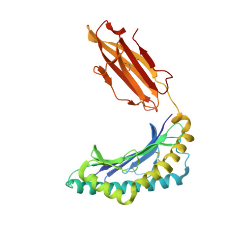
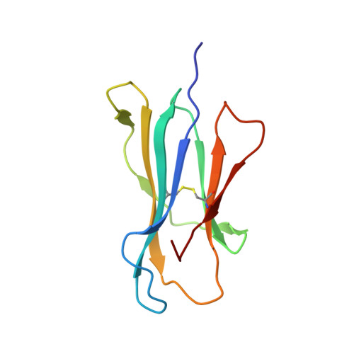
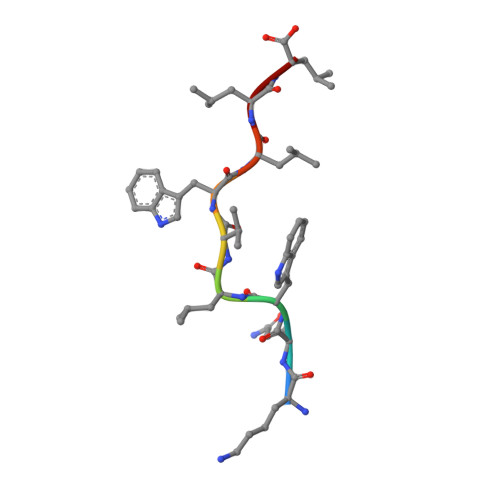
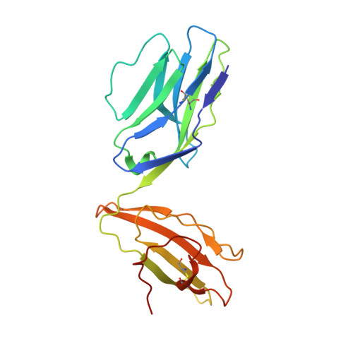
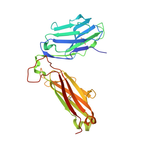
6UK2

Complex of T cell Receptor with HHAT Wild Type Peptide KQWLVWLLL Presented by HLA-A206


Experimental Data Snapshot

  • Method: X-RAY DIFFRACTION
  • Resolution: 3.14 Å
  • R-Value Free: 0.260 
  • R-Value Work: 0.229 
  • R-Value Observed: 0.231 

Starting Models: experimental
View more details

wwPDB Validation   3D Report Full Report

Currently 6UK2 does not have a validation slider image.


This is version 1.5 of the entry. See complete history


Literature

Structural dissimilarity from self drives neoepitope escape from immune tolerance.

Devlin, J.R.Alonso, J.A.Ayres, C.M.Keller, G.L.J.Bobisse, S.Vander Kooi, C.W.Coukos, G.Gfeller, D.Harari, A.Baker, B.M.

(2020) Nat Chem Biol 16: 1269-1276

  • DOI: https://doi.org/10.1038/s41589-020-0610-1
  • Primary Citation of Related Structures:  
    6UJO, 6UJQ, 6UK2, 6UK4

  • PubMed Abstract: 

    T-cell recognition of peptides incorporating nonsynonymous mutations, or neoepitopes, is a cornerstone of tumor immunity and forms the basis of new immunotherapy approaches including personalized cancer vaccines. Yet as they are derived from self-peptides, the means through which immunogenic neoepitopes overcome immune self-tolerance are often unclear. Here we show that a point mutation in a non-major histocompatibility complex anchor position induces structural and dynamic changes in an immunologically active ovarian cancer neoepitope. The changes pre-organize the peptide into a conformation optimal for recognition by a neoepitope-specific T-cell receptor, allowing the receptor to bind the neoepitope with high affinity and deliver potent T-cell signals. Our results emphasize the importance of structural and physical changes relative to self in neoepitope immunogenicity. Considered broadly, these findings can help explain some of the difficulties in identifying immunogenic neoepitopes from sequence alone and provide guidance for developing novel, neoepitope-based personalized therapies.


  • Organizational Affiliation

    Department of Chemistry and Biochemistry, University of Notre Dame, Notre Dame, IN, USA.


Macromolecules
Find similar proteins by:  (by identity cutoff)  |  3D Structure
Entity ID: 1
MoleculeChains Sequence LengthOrganismDetailsImage
MHC class I antigen276Homo sapiensMutation(s): 0 
Gene Names: HLA-A
UniProt
Find proteins for U5YJP1 (Homo sapiens)
Explore U5YJP1 
Go to UniProtKB:  U5YJP1
Entity Groups  
Sequence Clusters30% Identity50% Identity70% Identity90% Identity95% Identity100% Identity
UniProt GroupU5YJP1
Sequence Annotations
Expand
  • Reference Sequence
Find similar proteins by:  (by identity cutoff)  |  3D Structure
Entity ID: 2
MoleculeChains Sequence LengthOrganismDetailsImage
Beta-2-microglobulin100Homo sapiensMutation(s): 0 
Gene Names: B2MCDABP0092HDCMA22P
UniProt & NIH Common Fund Data Resources
Find proteins for P61769 (Homo sapiens)
Explore P61769 
Go to UniProtKB:  P61769
PHAROS:  P61769
GTEx:  ENSG00000166710 
Entity Groups  
Sequence Clusters30% Identity50% Identity70% Identity90% Identity95% Identity100% Identity
UniProt GroupP61769
Sequence Annotations
Expand
  • Reference Sequence

Find similar proteins by:  Sequence   |   3D Structure  

Entity ID: 3
MoleculeChains Sequence LengthOrganismDetailsImage
Protein-cysteine N-palmitoyltransferase HHAT9Homo sapiensMutation(s): 0 
EC: 2.3.1
UniProt & NIH Common Fund Data Resources
Find proteins for Q5VTY9 (Homo sapiens)
Explore Q5VTY9 
Go to UniProtKB:  Q5VTY9
PHAROS:  Q5VTY9
GTEx:  ENSG00000054392 
Entity Groups  
UniProt GroupQ5VTY9
Sequence Annotations
Expand
  • Reference Sequence
Find similar proteins by:  (by identity cutoff)  |  3D Structure
Entity ID: 4
MoleculeChains Sequence LengthOrganismDetailsImage
302 TIL T cell receptor alpha chain208Homo sapiensMutation(s): 0 
Entity Groups  
Sequence Clusters30% Identity50% Identity70% Identity90% Identity95% Identity100% Identity
Sequence Annotations
Expand
  • Reference Sequence
Find similar proteins by:  (by identity cutoff)  |  3D Structure
Entity ID: 5
MoleculeChains Sequence LengthOrganismDetailsImage
302 TIL T cell receptor beta chain245Homo sapiensMutation(s): 0 
Entity Groups  
Sequence Clusters30% Identity50% Identity70% Identity90% Identity95% Identity100% Identity
Sequence Annotations
Expand
  • Reference Sequence
Experimental Data & Validation

Experimental Data

  • Method: X-RAY DIFFRACTION
  • Resolution: 3.14 Å
  • R-Value Free: 0.260 
  • R-Value Work: 0.229 
  • R-Value Observed: 0.231 
  • Space Group: P 21 21 21
Unit Cell:
Length ( Å )Angle ( ˚ )
a = 64.357α = 90
b = 85.932β = 90
c = 240.728γ = 90
Software Package:
Software NamePurpose
PHENIXmodel building
PHENIXrefinement
HKL-2000data reduction
HKL-2000data scaling
PHASERphasing

Structure Validation

View Full Validation Report

Currently 6UK2 does not have a validation slider image.


Entry History & Funding Information

Deposition Data


Funding OrganizationLocationGrant Number
National Institutes of Health/National Institute of General Medical Sciences (NIH/NIGMS)United States--

Revision History  (Full details and data files)

  • Version 1.0: 2020-08-19
    Type: Initial release
  • Version 1.1: 2020-08-26
    Changes: Database references
  • Version 1.2: 2020-09-02
    Changes: Database references
  • Version 1.3: 2020-10-28
    Changes: Database references
  • Version 1.4: 2023-10-11
    Changes: Data collection, Database references, Refinement description
  • Version 1.5: 2024-10-30
    Changes: Structure summary