6UIH | pdb_00006uih

Crystal structure of the core domain from the GST-like protein GDAP1


Experimental Data Snapshot

  • Method: X-RAY DIFFRACTION
  • Resolution: 2.83 Å
  • R-Value Free: 
    0.224 (Depositor), 0.222 (DCC) 
  • R-Value Work: 
    0.211 (Depositor), 0.215 (DCC) 
  • R-Value Observed: 
    0.212 (Depositor) 

wwPDB Validation 3D Report Full Report

Validation slider image for 6UIH

This is version 1.2 of the entry. See complete history

Literature

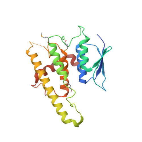
Structural and functional divergence of GDAP1 from the glutathione S-transferase superfamily.

Googins, M.R.Woghiren-Afegbua, A.O.Calderon, M.St Croix, C.M.Kiselyov, K.I.VanDemark, A.P.

(2020) FASEB J 34: 7192-7207

  • DOI: https://doi.org/10.1096/fj.202000110R
  • Primary Citation Related Structures: 
    6UIH

  • PubMed Abstract: 

    Mutations in ganglioside-induced differentiation-associated protein 1 (GDAP1) alter mitochondrial morphology and result in several subtypes of the inherited peripheral neuropathy Charcot-Marie-Tooth disease; however, the mechanism by which GDAP1 functions has remained elusive. GDAP1 contains primary sequence homology to the GST superfamily; however, the question of whether GDAP1 is an active GST has not been clearly resolved. Here, we present biochemical evidence, suggesting that GDAP1 has lost the ability to bind glutathione without a loss of substrate binding activity. We have revealed that the α-loop, located within the H-site motif is the primary determinant for substrate binding. Using structural data of GDAP1, we have found that critical residues and configurations in the G-site which canonically interact with glutathione are altered in GDAP1, rendering it incapable of binding glutathione. Last, we have found that the overexpression of GDAP1 in HeLa cells results in a mitochondrial phenotype which is distinct from oxidative stress-induced mitochondrial fragmentation. This phenotype is dependent on the presence of the transmembrane domain, as well as a unique hydrophobic domain that is not found in canonical GSTs. Together, we data point toward a non-enzymatic role for GDAP1, such as a sensor or receptor.


  • Organizational Affiliation
    • Department of Biological Sciences, University of Pittsburgh, Pittsburgh, PA, USA.

Macromolecule Content 

  • Total Structure Weight: 26.83 kDa 
  • Atom Count: 1,634 
  • Modeled Residue Count: 198 
  • Deposited Residue Count: 229 
  • Unique protein chains: 1

Macromolecules

Find similar proteins by:|  3D Structure
Entity ID: 1
MoleculeChains  Sequence LengthOrganismDetailsImage
Ganglioside-induced differentiation-associated protein 1229Mus musculusMutation(s): 0 
Gene Names: Gdap1
UniProt & NIH Common Fund Data Resources
Find proteins for O88741 (Mus musculus)
Explore O88741 
Go to UniProtKB:  O88741
IMPC:  MGI:1338002
Entity Groups
Sequence Clusters30% Identity50% Identity70% Identity90% Identity95% Identity100% Identity
UniProt GroupO88741
Sequence Annotations
Expand
Reference Sequence

Small Molecules

Modified Residues  1 Unique
IDChains TypeFormula2D DiagramParent
MSE
Query on MSE
A
L-PEPTIDE LINKINGC5 H11 N O2 SeMET

Experimental Data & Validation

Experimental Data

  • Method: X-RAY DIFFRACTION
  • Resolution: 2.83 Å
  • R-Value Free:  0.224 (Depositor), 0.222 (DCC) 
  • R-Value Work:  0.211 (Depositor), 0.215 (DCC) 
  • R-Value Observed: 0.212 (Depositor) 
Space Group: I 4 3 2
Unit Cell:
Length ( Å )Angle ( ˚ )
a = 196.598α = 90
b = 196.598β = 90
c = 196.598γ = 90
Software Package:
Software NamePurpose
Aimlessdata scaling
PHENIXrefinement
PDB_EXTRACTdata extraction
XDSdata reduction
AutoSolphasing

Structure Validation

View Full Validation Report


Entry History 

Deposition Data

Revision History  (Full details and data files)

  • Version 1.0: 2020-04-29
    Type: Initial release
  • Version 1.1: 2020-05-13
    Changes: Database references
  • Version 1.2: 2024-10-16
    Changes: Data collection, Database references, Structure summary