6UFI | pdb_00006ufi

W96Y Oxalate Decarboxylase (Bacillus subtilis)


Experimental Data Snapshot

  • Method: X-RAY DIFFRACTION
  • Resolution: 1.72 Å
  • R-Value Free: 
    0.166 (Depositor), 0.166 (DCC) 
  • R-Value Work: 
    0.149 (Depositor), 0.149 (DCC) 
  • R-Value Observed: 
    0.150 (Depositor) 

Starting Model: experimental
View more details

wwPDB Validation 3D Report Full Report

Validation slider image for 6UFI

This is version 1.4 of the entry. See complete history

Literature

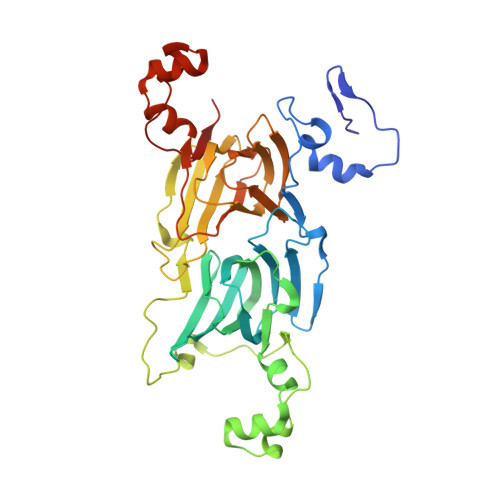
Oxalate decarboxylase uses electron hole hopping for catalysis.

Pastore, A.J.Teo, R.D.Montoya, A.Burg, M.J.Twahir, U.T.Bruner, S.D.Beratan, D.N.Angerhofer, A.

(2021) J Biological Chem 297: 100857-100857

  • DOI: https://doi.org/10.1016/j.jbc.2021.100857
  • Primary Citation Related Structures: 
    6TZP, 6UFI

  • PubMed Abstract: 

    The hexameric low-pH stress response enzyme oxalate decarboxylase catalyzes the decarboxylation of the oxalate mono-anion in the soil bacterium Bacillus subtilis. A single protein subunit contains two Mn-binding cupin domains, and catalysis depends on Mn(III) at the N-terminal site. The present study suggests a mechanistic function for the C-terminal Mn as an electron hole donor for the N-terminal Mn. The resulting spatial separation of the radical intermediates directs the chemistry toward decarboxylation of the substrate. A π-stacked tryptophan pair (W96/W274) links two neighboring protein subunits together, thus reducing the Mn-to-Mn distance from 25.9 Å (intrasubunit) to 21.5 Å (intersubunit). Here, we used theoretical analysis of electron hole-hopping paths through redox-active sites in the enzyme combined with site-directed mutagenesis and X-ray crystallography to demonstrate that this tryptophan pair supports effective electron hole hopping between the C-terminal Mn of one subunit and the N-terminal Mn of the other subunit through two short hops of ∼8.5 Å. Replacement of W96, W274, or both with phenylalanine led to a large reduction in catalytic efficiency, whereas replacement with tyrosine led to recovery of most of this activity. W96F and W96Y mutants share the wildtype tertiary structure. Two additional hole-hopping networks were identified leading from the Mn ions to the protein surface, potentially protecting the enzyme from high Mn oxidation states during turnover. Our findings strongly suggest that multistep hole-hopping transport between the two Mn ions is required for enzymatic function, adding to the growing examples of proteins that employ aromatic residues as hopping stations.


  • Organizational Affiliation
    • Department of Chemistry, University of Florida, Gainesville, Florida, USA.

Macromolecule Content 

  • Total Structure Weight: 43.89 kDa 
  • Atom Count: 3,620 
  • Modeled Residue Count: 377 
  • Deposited Residue Count: 385 
  • Unique protein chains: 1

Macromolecules

Find similar proteins by:|  3D Structure
Entity ID: 1
MoleculeChains  Sequence LengthOrganismDetailsImage
Cupin domain-containing protein385Bacillus subtilisMutation(s): 1 
Gene Names: B4417_3145ETL41_08750
EC: 4.1.1.2
UniProt
Find proteins for O34714 (Bacillus subtilis (strain 168))
Explore O34714 
Go to UniProtKB:  O34714
Entity Groups
Sequence Clusters30% Identity50% Identity70% Identity90% Identity95% Identity100% Identity
UniProt GroupO34714
Sequence Annotations
Expand
Reference Sequence

Small Molecules

Ligands 4 Unique
IDChains Name / Formula / InChI Key2D Diagram3D Interactions
GOL

Query on GOL


Download:Ideal Coordinates CCD File
D [auth A]GLYCEROL
C3 H8 O3
PEDCQBHIVMGVHV-UHFFFAOYSA-N
MN
(Subject of Investigation/LOI)

Query on MN


Download:Ideal Coordinates CCD File
B [auth A],
C [auth A]
MANGANESE (II) ION
Mn
WAEMQWOKJMHJLA-UHFFFAOYSA-N
CL

Query on CL


Download:Ideal Coordinates CCD File
E [auth A],
F [auth A]
CHLORIDE ION
Cl
VEXZGXHMUGYJMC-UHFFFAOYSA-M
NA

Query on NA


Download:Ideal Coordinates CCD File
G [auth A]SODIUM ION
Na
FKNQFGJONOIPTF-UHFFFAOYSA-N

Experimental Data & Validation

Experimental Data

  • Method: X-RAY DIFFRACTION
  • Resolution: 1.72 Å
  • R-Value Free:  0.166 (Depositor), 0.166 (DCC) 
  • R-Value Work:  0.149 (Depositor), 0.149 (DCC) 
  • R-Value Observed: 0.150 (Depositor) 
Space Group: H 3 2
Unit Cell:
Length ( Å )Angle ( ˚ )
a = 155.27α = 90
b = 155.27β = 90
c = 124.088γ = 120
Software Package:
Software NamePurpose
PHENIXrefinement
PDB_EXTRACTdata extraction
XDSdata reduction
Aimlessdata scaling
PHASERphasing

Structure Validation

View Full Validation Report


Entry History 

& Funding Information

Deposition Data


Funding OrganizationLocationGrant Number
National Science Foundation (NSF, United States)United States--

Revision History  (Full details and data files)

  • Version 1.0: 2020-09-30
    Type: Initial release
  • Version 1.1: 2020-10-07
    Changes: Data collection
  • Version 1.2: 2021-06-30
    Changes: Database references
  • Version 1.3: 2021-07-14
    Changes: Database references
  • Version 1.4: 2023-10-11
    Changes: Data collection, Database references, Refinement description