6UD5 | pdb_00006ud5

Crystal structure of human tryptophan 2,3-dioxygenase in complex with carbon monoxide and tryptophan


Experimental Data Snapshot

  • Method: X-RAY DIFFRACTION
  • Resolution: 2.05 Å
  • R-Value Free: 
    0.217 (Depositor), 0.228 (DCC) 
  • R-Value Work: 
    0.183 (Depositor) 
  • R-Value Observed: 
    0.185 (Depositor) 

Starting Model: experimental
View more details

wwPDB Validation 3D Report Full Report

Validation slider image for 6UD5

Ligand Structure Quality Assessment 


This is version 1.2 of the entry. See complete history

Literature

Conformational Plasticity in Human Heme-Based Dioxygenases.

Pham, K.N.Lewis-Ballester, A.Yeh, S.R.

(2021) J Am Chem Soc 143: 1836-1845

  • DOI: https://doi.org/10.1021/jacs.0c09970
  • Primary Citation Related Structures: 
    6UBP, 6UD5

  • PubMed Abstract: 

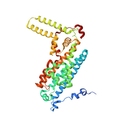
    Human indoleamine 2,3-dioxygenase 1 (hIDO1) and human tryptophan dioxygenase (hTDO) are two important heme proteins that degrade the essential amino acid, l-tryptophan (Trp), along the kynurenine pathway. The two enzymes share a similar active site structure and an analogous catalytic mechanism, but they exhibit a variety of distinct functional properties. Here we used carbon monoxide (CO) as a structural probe to interrogate how the functionalities of the two enzymes are encoded in their structures. With X-ray crystallography, we detected an unexpected photochemical intermediate trapped in a crystal of the hIDO1-CO-Trp complex, where CO is photolyzed from the heme iron by X-rays at cryogenic temperatures (100 K). The CO photolysis triggers a large-scale migration of the substrate Trp, as well as the photolyzed CO, from the active site to a temporary binding site, Sa*. It is accompanied by a large conformational change to an active site loop, JK-Loop C , despite the severely restricted protein motion under the frozen conditions, which highlights the remarkable conformational plasticity of the hIDO1 protein. Comparative studies of a crystal of the hTDO-CO-Trp complex show that CO and Trp remain bound in the active site under comparable X-ray illumination, indicating a much more rigid protein architecture. The data offer important new insights into the structure and function relationships of the heme-based dioxygenases and provide new guidelines for structure-based design of inhibitors targeting them.


  • Organizational Affiliation
    • Department of Physiology and Biophysics, Albert Einstein College of Medicine, The Bronx, New York 10461, United States.

Macromolecule Content 

  • Total Structure Weight: 184.94 kDa 
  • Atom Count: 12,705 
  • Modeled Residue Count: 1,394 
  • Deposited Residue Count: 1,520 
  • Unique protein chains: 1

Macromolecules

Find similar proteins by:|  3D Structure
Entity ID: 1
MoleculeChains  Sequence LengthOrganismDetailsImage
Tryptophan 2,3-dioxygenase
A, B, C, D
380Homo sapiensMutation(s): 0 
Gene Names: TDO2TDO
EC: 1.13.11.11
UniProt & NIH Common Fund Data Resources
Find proteins for P48775 (Homo sapiens)
Explore P48775 
Go to UniProtKB:  P48775
PHAROS:  P48775
GTEx:  ENSG00000151790 
Entity Groups
Sequence Clusters30% Identity50% Identity70% Identity90% Identity95% Identity100% Identity
UniProt GroupP48775
Sequence Annotations
Expand
Reference Sequence

Small Molecules

Ligands 3 Unique
IDChains Name / Formula / InChI Key2D Diagram3D Interactions
HEM

Query on HEM


Download:Ideal Coordinates CCD File
E [auth A],
I [auth B],
M [auth C],
Q [auth D]
PROTOPORPHYRIN IX CONTAINING FE
C34 H32 Fe N4 O4
KABFMIBPWCXCRK-RGGAHWMASA-L
TRP
(Subject of Investigation/LOI)

Query on TRP


Download:Ideal Coordinates CCD File
G [auth A]
H [auth A]
K [auth B]
L [auth B]
O [auth C]
G [auth A],
H [auth A],
K [auth B],
L [auth B],
O [auth C],
P [auth C],
S [auth D],
T [auth D]
TRYPTOPHAN
C11 H12 N2 O2
QIVBCDIJIAJPQS-VIFPVBQESA-N
CMO

Query on CMO


Download:Ideal Coordinates CCD File
F [auth A],
J [auth B],
N [auth C],
R [auth D]
CARBON MONOXIDE
C O
UGFAIRIUMAVXCW-UHFFFAOYSA-N

Experimental Data & Validation

Experimental Data

  • Method: X-RAY DIFFRACTION
  • Resolution: 2.05 Å
  • R-Value Free:  0.217 (Depositor), 0.228 (DCC) 
  • R-Value Work:  0.183 (Depositor) 
  • R-Value Observed: 0.185 (Depositor) 
Space Group: P 21 21 2
Unit Cell:
Length ( Å )Angle ( ˚ )
a = 144.725α = 90
b = 154.493β = 90
c = 88.306γ = 90
Software Package:
Software NamePurpose
REFMACrefinement
PDB_EXTRACTdata extraction
XDSdata reduction
SCALAdata scaling
PHASERphasing

Structure Validation

View Full Validation Report


Ligand Structure Quality Assessment 


Entry History 

& Funding Information

Deposition Data


Funding OrganizationLocationGrant Number
National Institutes of Health/National Institute of General Medical Sciences (NIH/NIGMS)United StatesGM115773
National Science Foundation (NSF, United States)United StatesCHE-1404929

Revision History  (Full details and data files)

  • Version 1.0: 2021-02-03
    Type: Initial release
  • Version 1.1: 2021-02-17
    Changes: Database references
  • Version 1.2: 2023-10-11
    Changes: Data collection, Database references, Refinement description