6UAK | pdb_00006uak

LahSb - C-terminal methyltransferase involved in RiPP biosynthesis


Experimental Data Snapshot

  • Method: X-RAY DIFFRACTION
  • Resolution: 2.01 Å
  • R-Value Free: 
    0.277 (Depositor), 0.280 (DCC) 
  • R-Value Work: 
    0.239 (Depositor), 0.241 (DCC) 
  • R-Value Observed: 
    0.240 (Depositor) 

wwPDB Validation 3D Report Full Report

Validation slider image for 6UAK

Ligand Structure Quality Assessment 


This is version 1.3 of the entry. See complete history

Literature

Characterization of a Dehydratase and Methyltransferase in the Biosynthesis of Ribosomally Synthesized and Post-translationally Modified Peptides in Lachnospiraceae.

Huo, L.Zhao, X.Acedo, J.Z.Estrada, P.Nair, S.K.van der Donk, W.A.

(2020) Chembiochem 21: 190-199

  • DOI: https://doi.org/10.1002/cbic.201900483
  • Primary Citation Related Structures: 
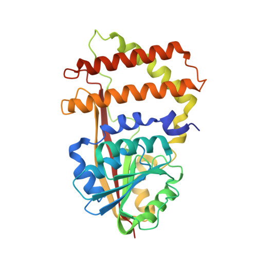
    6UAK

  • PubMed Abstract: 

    As a result of the exponential increase in genomic data, discovery of novel ribosomally synthesized and post-translationally modified peptide natural products (RiPPs) has progressed rapidly in the past decade. The lanthipeptides are a major subset of RiPPs. Through genome mining we identified a novel lanthipeptide biosynthetic gene cluster (lah) from Lachnospiraceae bacterium C6A11, an anaerobic bacterium that is a member of the human microbiota and which is implicated in the development of host disease states such as type 2 diabetes and resistance to Clostridium difficile colonization. The lah cluster encodes at least seven putative precursor peptides and multiple post-translational modification (PTM) enzymes. Two unusual class II lanthipeptide synthetases LahM1/M2 and a substrate-tolerant S-adenosyl-l-methionine (SAM)-dependent methyltransferase LahS B are biochemically characterized in this study. We also present the crystal structure of LahS B in complex with product S-adenosylhomocysteine. This study sets the stage for further exploration of the final products of the lah pathway as well as their potential physiological functions in human/animal gut microbiota.


  • Organizational Affiliation
    • Department of Chemistry and Howard Hughes Medical Institute, University of Illinois at Urbana-Champaign, 600 South Mathews Avenue, Urbana, IL, 61801, USA.

Macromolecule Content 

  • Total Structure Weight: 35.71 kDa 
  • Atom Count: 2,409 
  • Modeled Residue Count: 297 
  • Deposited Residue Count: 308 
  • Unique protein chains: 1

Macromolecules

Find similar proteins by:|  3D Structure
Entity ID: 1
MoleculeChains  Sequence LengthOrganismDetailsImage
SAM dependent methyltransferase LahSB308Lachnospiraceae bacterium C6A11Mutation(s): 0 
Gene Names: DEO87_04690

Small Molecules

Ligands 1 Unique
IDChains Name / Formula / InChI Key2D Diagram3D Interactions
SAH

Query on SAH


Download:Ideal Coordinates CCD File
B [auth A]S-ADENOSYL-L-HOMOCYSTEINE
C14 H20 N6 O5 S
ZJUKTBDSGOFHSH-WFMPWKQPSA-N

Experimental Data & Validation

Experimental Data

  • Method: X-RAY DIFFRACTION
  • Resolution: 2.01 Å
  • R-Value Free:  0.277 (Depositor), 0.280 (DCC) 
  • R-Value Work:  0.239 (Depositor), 0.241 (DCC) 
  • R-Value Observed: 0.240 (Depositor) 
Space Group: P 32 2 1
Unit Cell:
Length ( Å )Angle ( ˚ )
a = 115.729α = 90
b = 115.729β = 90
c = 71.26γ = 120
Software Package:
Software NamePurpose
REFMACrefinement
PDB_EXTRACTdata extraction
XDSdata reduction
pointlessdata scaling
SHARPphasing

Structure Validation

View Full Validation Report


Ligand Structure Quality Assessment 


Entry History 

& Funding Information

Deposition Data


Funding OrganizationLocationGrant Number
National Institutes of Health/National Institute of General Medical Sciences (NIH/NIGMS)United States--

Revision History  (Full details and data files)

  • Version 1.0: 2019-12-04
    Type: Initial release
  • Version 1.1: 2020-01-01
    Changes: Author supporting evidence
  • Version 1.2: 2020-02-05
    Changes: Database references
  • Version 1.3: 2024-03-13
    Changes: Data collection, Database references