6U7T | pdb_00006u7t

MutY adenine glycosylase bound to DNA containing a transition state analog (1N) paired with d(8-oxo-G)


Experimental Data Snapshot

  • Method: X-RAY DIFFRACTION
  • Resolution: 2.00 Å
  • R-Value Free: 
    0.270 (Depositor), 0.270 (DCC) 
  • R-Value Work: 
    0.261 (Depositor), 0.262 (DCC) 
  • R-Value Observed: 
    0.262 (Depositor) 

Starting Model: experimental
View more details

wwPDB Validation 3D Report Full Report

Validation slider image for 6U7T

This is version 2.2 of the entry. See complete history

Literature

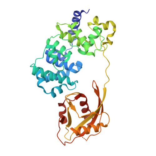
Structural Basis for Finding OG Lesions and Avoiding Undamaged G by the DNA Glycosylase MutY.

Russelburg, L.P.O'Shea Murray, V.L.Demir, M.Knutsen, K.R.Sehgal, S.L.Cao, S.David, S.S.Horvath, M.P.

(2020) ACS Chem Biol 15: 93-102

  • DOI: https://doi.org/10.1021/acschembio.9b00639
  • Primary Citation Related Structures: 
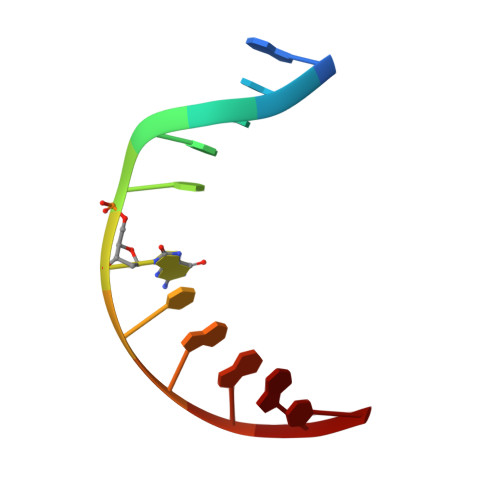
    6Q0C, 6U7T

  • PubMed Abstract: 

    The adenine glycosylase MutY selectively initiates repair of OG:A lesions and, by comparison, avoids G:A mispairs. The ability to distinguish these closely related substrates relies on the C-terminal domain of MutY, which structurally resembles MutT. To understand the mechanism for substrate specificity, we crystallized MutY in complex with DNA containing G across from the high-affinity azaribose transition state analogue. Our structure shows that G is accommodated by the OG site and highlights the role of a serine residue in OG versus G discrimination. The functional significance of Ser308 and its neighboring residues was evaluated by mutational analysis, revealing the critical importance of a β loop in the C-terminal domain for mutation suppression in cells, and biochemical performance in vitro . This loop comprising residues Phe307, Ser308, and His309 ( Geobacillus stearothermophilus sequence positions) is conserved in MutY but absent in MutT and other DNA repair enzymes and may therefore serve as a MutY-specific target exploitable by chemical biological probes.


  • Organizational Affiliation
    • School of Biological Sciences , University of Utah , 257 South 1400 East , Salt Lake City , Utah 84112 , United States.

Macromolecule Content 

  • Total Structure Weight: 48.86 kDa 
  • Atom Count: 3,252 
  • Modeled Residue Count: 370 
  • Deposited Residue Count: 388 
  • Unique protein chains: 1
  • Unique nucleic acid chains: 2

Macromolecules


Find similar proteins by:|  3D Structure
Entity ID: 1
MoleculeChains  Sequence LengthOrganismDetailsImage
Adenine DNA glycosylase366Geobacillus stearothermophilusMutation(s): 2 
Gene Names: mutY
EC: 3.2.2.31
UniProt
Find proteins for P83847 (Geobacillus stearothermophilus)
Explore P83847 
Go to UniProtKB:  P83847
Entity Groups
Sequence Clusters30% Identity50% Identity70% Identity90% Identity95% Identity100% Identity
UniProt GroupP83847
Sequence Annotations
Expand
Reference Sequence
Find similar nucleic acids by:  Sequence
Entity ID: 2
MoleculeChains LengthOrganismImage
DNA (5'-D(*AP*AP*GP*AP*CP*(8OG)P*TP*GP*GP*AP*C)-3')11synthetic construct
Sequence Annotations
Expand
Reference Sequence
Find similar nucleic acids by:  Sequence
Entity ID: 3
MoleculeChains LengthOrganismImage
DNA (5'-D(P*GP*TP*CP*CP*AP*(NR1)P*GP*TP*CP*T)-3')11synthetic construct
Sequence Annotations
Expand
Reference Sequence

Experimental Data & Validation

Experimental Data

  • Method: X-RAY DIFFRACTION
  • Resolution: 2.00 Å
  • R-Value Free:  0.270 (Depositor), 0.270 (DCC) 
  • R-Value Work:  0.261 (Depositor), 0.262 (DCC) 
  • R-Value Observed: 0.262 (Depositor) 
Space Group: P 21 21 21
Unit Cell:
Length ( Å )Angle ( ˚ )
a = 37.8α = 90
b = 86.49β = 90
c = 140.58γ = 90
Software Package:
Software NamePurpose
XSCALEdata scaling
PHENIXrefinement
PDB_EXTRACTdata extraction
XDSdata reduction
PHASERphasing

Structure Validation

View Full Validation Report


Entry History 

& Funding Information

Deposition Data


Funding OrganizationLocationGrant Number
National Science Foundation (NSF, United States)United States1608934
National Science Foundation (NSF, United States)United States1610721

Revision History  (Full details and data files)

  • Version 1.0: 2019-10-02
    Type: Initial release
  • Version 1.1: 2019-11-13
    Changes: Database references
  • Version 1.2: 2019-11-20
    Changes: Derived calculations
  • Version 1.3: 2019-11-27
    Changes: Author supporting evidence
  • Version 2.0: 2019-12-25
    Changes: Atomic model, Database references
  • Version 2.1: 2020-01-29
    Changes: Database references
  • Version 2.2: 2023-10-11
    Changes: Data collection, Database references, Derived calculations, Refinement description