6U7M | pdb_00006u7m

Cryo-EM Structure of Helical Lipoprotein Lipase


Experimental Data Snapshot

  • Method: ELECTRON MICROSCOPY
  • Resolution: 3.80 Å
  • Aggregation State: FILAMENT 
  • Reconstruction Method: HELICAL 

Starting Model: experimental
View more details

wwPDB Validation 3D Report Full Report

Validation slider image for 6U7M

This is version 1.4 of the entry. See complete history

Literature

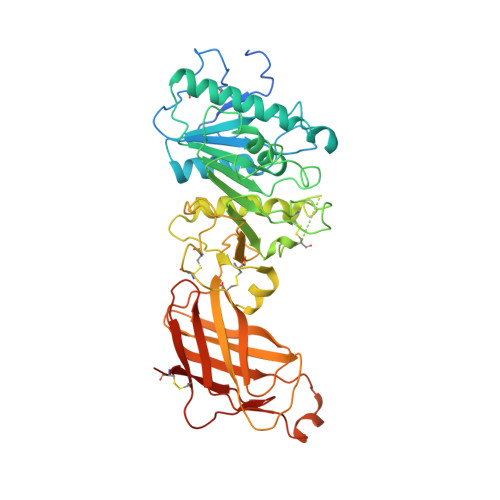
The structure of helical lipoprotein lipase reveals an unexpected twist in lipase storage.

Gunn, K.H.Roberts, B.S.Wang, F.Strauss, J.D.Borgnia, M.J.Egelman, E.H.Neher, S.B.

(2020) Proc Natl Acad Sci U S A 117: 10254-10264

  • DOI: https://doi.org/10.1073/pnas.1916555117
  • Primary Citation Related Structures: 
    6U7M

  • PubMed Abstract: 

    Lipases are enzymes necessary for the proper distribution and utilization of lipids in the human body. Lipoprotein lipase (LPL) is active in capillaries, where it plays a crucial role in preventing dyslipidemia by hydrolyzing triglycerides from packaged lipoproteins. Thirty years ago, the existence of a condensed and inactive LPL oligomer was proposed. Although recent work has shed light on the structure of the LPL monomer, the inactive oligomer remained opaque. Here we present a cryo-EM reconstruction of a helical LPL oligomer at 3.8-Å resolution. Helix formation is concentration-dependent, and helices are composed of inactive dihedral LPL dimers. Heparin binding stabilizes LPL helices, and the presence of substrate triggers helix disassembly. Superresolution fluorescent microscopy of endogenous LPL revealed that LPL adopts a filament-like distribution in vesicles. Mutation of one of the helical LPL interaction interfaces causes loss of the filament-like distribution. Taken together, this suggests that LPL is condensed into its inactive helical form for storage in intracellular vesicles.


  • Organizational Affiliation
    • Department of Biochemistry and Biophysics, University of North Carolina, Chapel Hill, NC 27599.

Macromolecule Content 

  • Total Structure Weight: 1,603.46 kDa 
  • Atom Count: 99,420 
  • Modeled Residue Count: 12,510 
  • Deposited Residue Count: 14,340 
  • Unique protein chains: 1

Macromolecules

Find similar proteins by:|  3D Structure
Entity ID: 1
MoleculeChains  Sequence LengthOrganismDetailsImage
Lipoprotein lipase478Bos taurusMutation(s): 0 
EC: 3.1.1.34 (PDB Primary Data), 3.1.1.32 (UniProt)
Membrane Entity: Yes 
UniProt
Find proteins for P11151 (Bos taurus)
Explore P11151 
Go to UniProtKB:  P11151
Entity Groups
Sequence Clusters30% Identity50% Identity70% Identity90% Identity95% Identity100% Identity
UniProt GroupP11151
Sequence Annotations
Expand
Reference Sequence

Experimental Data & Validation

Experimental Data

  • Method: ELECTRON MICROSCOPY
  • Resolution: 3.80 Å
  • Aggregation State: FILAMENT 
  • Reconstruction Method: HELICAL 
EM Software:
TaskSoftware PackageVersion
RECONSTRUCTIONRELION3.0
MODEL REFINEMENTPHENIX

Structure Validation

View Full Validation Report


Entry History 

& Funding Information

Deposition Data


Funding OrganizationLocationGrant Number
National Institutes of Health/National Human Genome Research Institute (NIH/NHGRI)United StatesR01-HL12565
National Institutes of Health/National Human Genome Research Institute (NIH/NHGRI)United StatesR35-GM122510

Revision History  (Full details and data files)

  • Version 1.0: 2020-04-08
    Type: Initial release
  • Version 1.1: 2020-05-06
    Changes: Database references
  • Version 1.2: 2020-05-27
    Changes: Database references
  • Version 1.3: 2024-11-20
    Changes: Data collection, Database references, Refinement description, Structure summary
  • Version 1.4: 2025-05-21
    Changes: Data collection