6U61

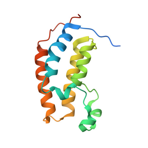
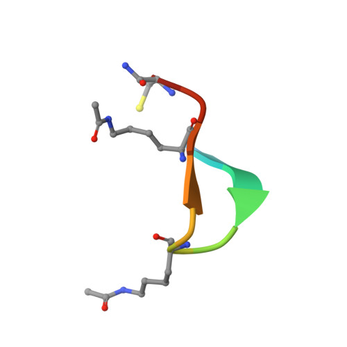
BRD2-BD1 in complex with the cyclic peptide 3.1_3


Experimental Data Snapshot

  • Method: X-RAY DIFFRACTION
  • Resolution: 2.29 Å
  • R-Value Free: 0.263 
  • R-Value Work: 0.222 
  • R-Value Observed: 0.224 

Starting Model: experimental
View more details

wwPDB Validation   3D Report Full Report


This is version 1.4 of the entry. See complete history


Literature

Cyclic peptides can engage a single binding pocket through highly divergent modes.

Patel, K.Walport, L.J.Walshe, J.L.Solomon, P.D.Low, J.K.K.Tran, D.H.Mouradian, K.S.Silva, A.P.G.Wilkinson-White, L.Norman, A.Franck, C.Matthews, J.M.Guss, J.M.Payne, R.J.Passioura, T.Suga, H.Mackay, J.P.

(2020) Proc Natl Acad Sci U S A 117: 26728-26738

  • DOI: https://doi.org/10.1073/pnas.2003086117
  • Primary Citation of Related Structures:  
    6U4A, 6U61, 6U6K, 6U6L, 6U71, 6U72, 6U74, 6U8G, 6U8H, 6U8I, 6U8M, 6ULP, 6ULQ, 6ULT, 6ULV

  • PubMed Abstract: 

    Cyclic peptide library screening technologies show immense promise for identifying drug leads and chemical probes for challenging targets. However, the structural and functional diversity encoded within such libraries is largely undefined. We have systematically profiled the affinity, selectivity, and structural features of library-derived cyclic peptides selected to recognize three closely related targets: the acetyllysine-binding bromodomain proteins BRD2, -3, and -4. We report affinities as low as 100 pM and specificities of up to 10 6 -fold. Crystal structures of 13 peptide-bromodomain complexes reveal remarkable diversity in both structure and binding mode, including both α-helical and β-sheet structures as well as bivalent binding modes. The peptides can also exhibit a high degree of structural preorganization. Our data demonstrate the enormous potential within these libraries to provide diverse binding modes against a single target, which underpins their capacity to yield highly potent and selective ligands.


  • Organizational Affiliation

    School of Life and Environmental Sciences, The University of Sydney, Sydney, NSW 2006, Australia.


Macromolecules
Find similar proteins by:  (by identity cutoff)  |  3D Structure
Entity ID: 1
MoleculeChains Sequence LengthOrganismDetailsImage
Bromodomain-containing protein 2
A, B
136Homo sapiensMutation(s): 0 
Gene Names: BRD2
UniProt & NIH Common Fund Data Resources
Find proteins for P25440 (Homo sapiens)
Explore P25440 
Go to UniProtKB:  P25440
PHAROS:  P25440
GTEx:  ENSG00000204256 
Entity Groups  
Sequence Clusters30% Identity50% Identity70% Identity90% Identity95% Identity100% Identity
UniProt GroupP25440
Sequence Annotations
Expand
  • Reference Sequence

Find similar proteins by:  Sequence   |   3D Structure  

Entity ID: 2
MoleculeChains Sequence LengthOrganismDetailsImage
cyclic peptide 3.1_3C [auth E],
D
13synthetic constructMutation(s): 0 
Entity Groups  
Sequence Clusters30% Identity50% Identity70% Identity90% Identity95% Identity100% Identity
Sequence Annotations
Expand
  • Reference Sequence
Experimental Data & Validation

Experimental Data

  • Method: X-RAY DIFFRACTION
  • Resolution: 2.29 Å
  • R-Value Free: 0.263 
  • R-Value Work: 0.222 
  • R-Value Observed: 0.224 
  • Space Group: C 2 2 21
Unit Cell:
Length ( Å )Angle ( ˚ )
a = 69.538α = 90
b = 86.752β = 90
c = 115.48γ = 90
Software Package:
Software NamePurpose
PHENIXrefinement
XDSdata reduction
Aimlessdata scaling
PHASERphasing

Structure Validation

View Full Validation Report


Entry History & Funding Information

Deposition Data


Funding OrganizationLocationGrant Number
National Health and Medical Research Council (NHMRC, Australia)AustraliaAPP1161623

Revision History  (Full details and data files)

  • Version 1.0: 2020-08-19
    Type: Initial release
  • Version 1.1: 2021-03-03
    Changes: Database references
  • Version 1.2: 2023-10-11
    Changes: Data collection, Database references, Refinement description
  • Version 1.3: 2023-11-15
    Changes: Data collection
  • Version 1.4: 2024-11-06
    Changes: Structure summary