6U3O | pdb_00006u3o

JR51 DQ2-p.aeru-alpha2a complex


Experimental Data Snapshot

  • Method: X-RAY DIFFRACTION
  • Resolution: 2.74 Å
  • R-Value Free: 
    0.230 (DCC) 

wwPDB Validation 3D Report Full Report

Validation slider image for 6U3O

This is version 2.1 of the entry. See complete history

Literature

T cell receptor cross-reactivity between gliadin and bacterial peptides in celiac disease.

Petersen, J.Ciacchi, L.Tran, M.T.Loh, K.L.Kooy-Winkelaar, Y.Croft, N.P.Hardy, M.Y.Chen, Z.McCluskey, J.Anderson, R.P.Purcell, A.W.Tye-Din, J.A.Koning, F.Reid, H.H.Rossjohn, J.

(2020) Nat Struct Mol Biol 27: 49-61

  • DOI: https://doi.org/10.1038/s41594-019-0353-4
  • Primary Citation Related Structures: 
    6U3M, 6U3N, 6U3O

  • PubMed Abstract: 

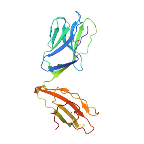
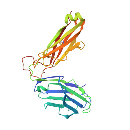
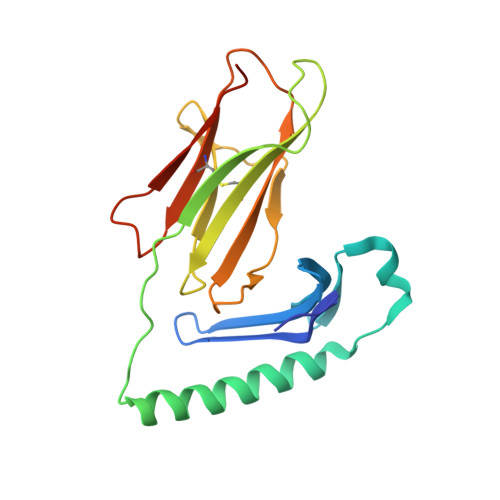
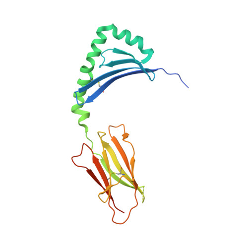
    The human leukocyte antigen (HLA) locus is strongly associated with T cell-mediated autoimmune disorders. HLA-DQ2.5-mediated celiac disease (CeD) is triggered by the ingestion of gluten, although the relative roles of genetic and environmental risk factors in CeD is unclear. Here we identify microbially derived mimics of gliadin epitopes and a parental bacterial protein that is naturally processed by antigen-presenting cells and activated gliadin reactive HLA-DQ2.5-restricted T cells derived from CeD patients. Crystal structures of T cell receptors in complex with HLA-DQ2.5 bound to two distinct bacterial peptides demonstrate that molecular mimicry underpins cross-reactivity toward the gliadin epitopes. Accordingly, gliadin reactive T cells involved in CeD pathogenesis cross-react with ubiquitous bacterial peptides, thereby suggesting microbial exposure as a potential environmental factor in CeD.


  • Organizational Affiliation
    • Infection and Immunity Program and The Department of Biochemistry and Molecular Biology, Biomedicine Discovery Institute Monash University, Clayton, Victoria, Australia.

Macromolecule Content 

  • Total Structure Weight: 194.92 kDa 
  • Atom Count: 12,888 
  • Modeled Residue Count: 1,622 
  • Deposited Residue Count: 1,726 
  • Unique protein chains: 5

Macromolecules

Find similar proteins by:|  3D Structure
Entity ID: 1
MoleculeChains  Sequence LengthOrganismDetailsImage
T-CELL RECEPTOR, JR5.1 alphaA [auth G],
C [auth A]
202Homo sapiensMutation(s): 0 
Find similar proteins by:|  3D Structure
Entity ID: 2
MoleculeChains  Sequence LengthOrganismDetailsImage
T-CELL RECEPTOR, JR5.1 betaB [auth H],
D [auth B]
244Homo sapiensMutation(s): 0 
Find similar proteins by:|  3D Structure
Entity ID: 3
MoleculeChains  Sequence LengthOrganismDetailsImage
MHC class II HLA-DQ-alpha chainE [auth C],
G [auth E]
191Homo sapiensMutation(s): 0 
Gene Names: HLA-DQA1
UniProt
Find proteins for O19705 (Homo sapiens)
Explore O19705 
Go to UniProtKB:  O19705
Entity Groups
Sequence Clusters30% Identity50% Identity70% Identity90% Identity95% Identity100% Identity
UniProt GroupO19705
Glycosylation
Glycosylation Sites: 1
Sequence Annotations
Expand
Reference Sequence
Find similar proteins by:|  3D Structure
Entity ID: 4
MoleculeChains  Sequence LengthOrganismDetailsImage
MHC class II HLA-DQ-beta-1F [auth D],
H [auth F]
206Homo sapiensMutation(s): 0 
Gene Names: HLA-DQB1
UniProt
Find proteins for O19712 (Homo sapiens)
Explore O19712 
Go to UniProtKB:  O19712
Entity Groups
Sequence Clusters30% Identity50% Identity70% Identity90% Identity95% Identity100% Identity
UniProt GroupO19712
Sequence Annotations
Expand
Reference Sequence
Find similar proteins by:  Sequence   |   3D Structure  
Entity ID: 5
MoleculeChains  Sequence LengthOrganismDetailsImage
Peptide
I, J
20Pseudomonas aeruginosaMutation(s): 0 
UniProt
Find proteins for Q9HX82 (Pseudomonas aeruginosa (strain ATCC 15692 / DSM 22644 / CIP 104116 / JCM 14847 / LMG 12228 / 1C / PRS 101 / PAO1))
Explore Q9HX82 
Go to UniProtKB:  Q9HX82
Entity Groups
Sequence Clusters30% Identity50% Identity70% Identity90% Identity95% Identity100% Identity
UniProt GroupQ9HX82
Sequence Annotations
Expand
Reference Sequence

Oligosaccharides

Help  
Entity ID: 6
MoleculeChains Length2D Diagram GlycosylationD Interactions
2-acetamido-2-deoxy-beta-D-glucopyranose-(1-4)-2-acetamido-2-deoxy-beta-D-glucopyranose
K, L
2N-Glycosylation
Glycosylation Resources
GlyTouCan: G42666HT
GlyCosmos: G42666HT
GlyGen: G42666HT

Experimental Data & Validation

Experimental Data

  • Method: X-RAY DIFFRACTION
  • Resolution: 2.74 Å
  • R-Value Free:  0.230 (DCC) 
Space Group: P 1 21 1
Unit Cell:
Length ( Å )Angle ( ˚ )
a = 69.057α = 90
b = 157.65β = 96.53
c = 106.1γ = 90
Software Package:
Software NamePurpose
PHENIXrefinement
XDSdata reduction
Aimlessdata scaling
PHASERphasing

Structure Validation

View Full Validation Report


Entry History 

& Funding Information

Deposition Data


Funding OrganizationLocationGrant Number
Australian Research Council (ARC)AustraliaCE140100011

Revision History  (Full details and data files)

  • Version 1.0: 2019-12-18
    Type: Initial release
  • Version 1.1: 2020-07-01
    Changes: Data collection, Database references
  • Version 2.0: 2020-07-29
    Type: Remediation
    Reason: Carbohydrate remediation
    Changes: Atomic model, Data collection, Derived calculations, Structure summary
  • Version 2.1: 2024-10-30
    Changes: Data collection, Database references, Structure summary