6U3L | pdb_00006u3l

Crystal structure of Hemerythrin HHE cation binding domain-containing protein: Rv2633c homolog from Mycobacterium kansasii


Experimental Data Snapshot

  • Method: X-RAY DIFFRACTION
  • Resolution: 1.75 Å
  • R-Value Free: 
    0.218 (Depositor), 0.221 (DCC) 
  • R-Value Work: 
    0.178 (Depositor), 0.180 (DCC) 
  • R-Value Observed: 
    0.182 (Depositor) 

wwPDB Validation 3D Report Full Report

Validation slider image for 6U3L

This is version 1.2 of the entry. See complete history

Literature

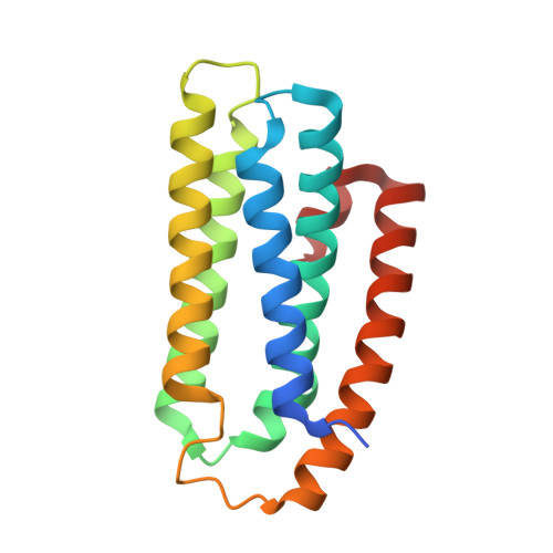
Crystal structure of a hemerythrin-like protein from Mycobacterium kansasii and homology model of the orthologous Rv2633c protein of M. tuberculosis.

Ma, Z.Abendroth, J.Buchko, G.W.Rohde, K.H.Davidson, V.L.

(2020) Biochem J 477: 567-581

  • DOI: https://doi.org/10.1042/BCJ20190827
  • Primary Citation Related Structures: 
    6Q09, 6U3L

  • PubMed Abstract: 

    Pathogenic and opportunistic mycobacteria have a distinct class of non-heme di-iron hemerythrin-like proteins (HLPs). The first to be isolated was the Rv2633c protein, which plays a role in infection by Mycobacterium tuberculosis (Mtb), but could not be crystallized. This work presents the first crystal structure of an ortholog of Rv2633c, the mycobacterial HLP from Mycobacterium kansasii (Mka). This structure differs from those of hemerythrins and other known HLPs. It consists of five α-helices, whereas all other HLP domains have four. In contrast with other HLPs, the HLP domain is not fused to an additional protein domain. The residues ligating and surrounding the di-iron site are also unique among HLPs. Notably, a tyrosine occupies the position normally held by one of the histidine ligands in hemerythrin. This structure was used to construct a homology model of Rv2633c. The structure of five α-helices is conserved and the di-iron site ligands are identical in Rv2633c. Two residues near the ends of helices in the Mka HLP structure are replaced with prolines in the Rv2633c model. This may account for structural perturbations that decrease the solubility of Rv2633c relative to Mka HLP. Clusters of residues that differ in charge or polarity between Rv2633c and Mka HLP that point outward from the helical core could reflect a specificity for potential differential interactions with other protein partners in vivo, which are related to function. The Mka HLP exhibited weaker catalase activity than Rv2633c. Evidence was obtained for the interaction of Mka HLP irons with nitric oxide.


  • Organizational Affiliation
    • Burnett School of Biomedical Sciences, College of Medicine, University of Central Florida, Orlando, FL 32827, U.S.A.

Macromolecule Content 

  • Total Structure Weight: 19.6 kDa 
  • Atom Count: 1,420 
  • Modeled Residue Count: 162 
  • Deposited Residue Count: 170 
  • Unique protein chains: 1

Macromolecules

Find similar proteins by:|  3D Structure
Entity ID: 1
MoleculeChains  Sequence LengthOrganismDetailsImage
Hemerythrin HHE cation binding domain protein170Mycobacterium kansasiiMutation(s): 1 
Gene Names: BZL29_7639
UniProt
Find proteins for A0A1V3WIE5 (Mycobacterium kansasii)
Explore A0A1V3WIE5 
Go to UniProtKB:  A0A1V3WIE5
Entity Groups
Sequence Clusters30% Identity50% Identity70% Identity90% Identity95% Identity100% Identity
UniProt GroupA0A1V3WIE5
Sequence Annotations
Expand
Reference Sequence

Small Molecules

Ligands 1 Unique
IDChains Name / Formula / InChI Key2D Diagram3D Interactions
EDO

Query on EDO


Download:Ideal Coordinates CCD File
B [auth A]1,2-ETHANEDIOL
C2 H6 O2
LYCAIKOWRPUZTN-UHFFFAOYSA-N

Experimental Data & Validation

Experimental Data

  • Method: X-RAY DIFFRACTION
  • Resolution: 1.75 Å
  • R-Value Free:  0.218 (Depositor), 0.221 (DCC) 
  • R-Value Work:  0.178 (Depositor), 0.180 (DCC) 
  • R-Value Observed: 0.182 (Depositor) 
Space Group: P 31 2 1
Unit Cell:
Length ( Å )Angle ( ˚ )
a = 52.05α = 90
b = 52.05β = 90
c = 104.7γ = 120
Software Package:
Software NamePurpose
XDSdata reduction
XSCALEdata scaling
PHENIXrefinement
PDB_EXTRACTdata extraction
PHASERphasing
PARROTphasing
ARP/wARPmodel building
Cootmodel building

Structure Validation

View Full Validation Report


Entry History 

Deposition Data

Revision History  (Full details and data files)

  • Version 1.0: 2020-01-22
    Type: Initial release
  • Version 1.1: 2020-02-12
    Changes: Database references
  • Version 1.2: 2024-03-13
    Changes: Data collection, Database references