6U2W

EM structure of MPEG-1(L425K) pre-pore complex bound to liposome


Experimental Data Snapshot

  • Method: ELECTRON MICROSCOPY
  • Resolution: 3.63 Å
  • Aggregation State: PARTICLE 
  • Reconstruction Method: SINGLE PARTICLE 

wwPDB Validation   3D Report Full Report


This is version 2.0 of the entry. See complete history


Literature

The cryo-EM structure of the acid activatable pore-forming immune effector Macrophage-expressed gene 1.

Pang, S.S.Bayly-Jones, C.Radjainia, M.Spicer, B.A.Law, R.H.P.Hodel, A.W.Parsons, E.S.Ekkel, S.M.Conroy, P.J.Ramm, G.Venugopal, H.Bird, P.I.Hoogenboom, B.W.Voskoboinik, I.Gambin, Y.Sierecki, E.Dunstone, M.A.Whisstock, J.C.

(2019) Nat Commun 10: 4288-4288

  • DOI: https://doi.org/10.1038/s41467-019-12279-2
  • Primary Citation of Related Structures:  
    6U23, 6U2J, 6U2K, 6U2L, 6U2W

  • PubMed Abstract: 

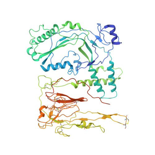
    Macrophage-expressed gene 1 (MPEG1/Perforin-2) is a perforin-like protein that functions within the phagolysosome to damage engulfed microbes. MPEG1 is thought to form pores in target membranes, however, its mode of action remains unknown. We use cryo-Electron Microscopy (cryo-EM) to determine the 2.4 Å structure of a hexadecameric assembly of MPEG1 that displays the expected features of a soluble prepore complex. We further discover that MPEG1 prepore-like assemblies can be induced to perforate membranes through acidification, such as would occur within maturing phagolysosomes. We next solve the 3.6 Å cryo-EM structure of MPEG1 in complex with liposomes. These data reveal that a multi-vesicular body of 12 kDa (MVB12)-associated β-prism (MABP) domain binds membranes such that the pore-forming machinery of MPEG1 is oriented away from the bound membrane. This unexpected mechanism of membrane interaction suggests that MPEG1 remains bound to the phagolysosome membrane while simultaneously forming pores in engulfed bacterial targets.


  • Organizational Affiliation

    ARC Centre of Excellence in Advanced Molecular Imaging, Monash University, Melbourne, VIC, 3800, Australia.


Macromolecules
Find similar proteins by:  (by identity cutoff)  |  3D Structure
Entity ID: 1
MoleculeChains Sequence LengthOrganismDetailsImage
Macrophage-expressed gene 1 protein
A, B, C, D, E
A, B, C, D, E, F, G, H, I, J, K, L, M, N, O, P
642Homo sapiensMutation(s): 1 
Gene Names: MPEG1
Membrane Entity: Yes 
UniProt & NIH Common Fund Data Resources
Find proteins for Q2M385 (Homo sapiens)
Explore Q2M385 
Go to UniProtKB:  Q2M385
PHAROS:  Q2M385
GTEx:  ENSG00000197629 
Entity Groups  
Sequence Clusters30% Identity50% Identity70% Identity90% Identity95% Identity100% Identity
UniProt GroupQ2M385
Sequence Annotations
Expand
  • Reference Sequence
Oligosaccharides

Help

Entity ID: 2
MoleculeChains Length2D Diagram Glycosylation3D Interactions
2-acetamido-2-deoxy-beta-D-glucopyranose-(1-4)-2-acetamido-2-deoxy-beta-D-glucopyranose
AA [auth a],
BA [auth b],
CA [auth c],
DA [auth d],
EA [auth e],
AA [auth a],
BA [auth b],
CA [auth c],
DA [auth d],
EA [auth e],
FA [auth f],
GA [auth g],
HA [auth h],
IA [auth i],
JA [auth j],
KA [auth k],
LA [auth l],
MA [auth m],
NA [auth n],
OA [auth o],
PA [auth p],
Q,
QA [auth q],
R,
RA [auth r],
S,
SA [auth s],
T,
TA [auth t],
U,
UA [auth u],
V,
VA [auth v],
W,
X,
Y,
Z
2N-Glycosylation
Glycosylation Resources
GlyTouCan:  G42666HT
GlyCosmos:  G42666HT
GlyGen:  G42666HT
Experimental Data & Validation

Experimental Data

  • Method: ELECTRON MICROSCOPY
  • Resolution: 3.63 Å
  • Aggregation State: PARTICLE 
  • Reconstruction Method: SINGLE PARTICLE 

Structure Validation

View Full Validation Report


Entry History & Funding Information

Deposition Data


Funding OrganizationLocationGrant Number
Australian Research Council (ARC)Australia--

Revision History  (Full details and data files)

  • Version 1.0: 2019-09-25
    Type: Initial release
  • Version 1.1: 2019-10-02
    Changes: Data collection, Database references
  • Version 1.2: 2020-01-15
    Changes: Author supporting evidence
  • Version 2.0: 2020-07-29
    Type: Remediation
    Reason: Carbohydrate remediation
    Changes: Atomic model, Data collection, Derived calculations, Structure summary