6U0K | pdb_00006u0k

TTBK2 kinase domain in complex with Compound 1


Experimental Data Snapshot

  • Method: X-RAY DIFFRACTION
  • Resolution: 1.74 Å
  • R-Value Free: 
    0.233 (Depositor), 0.235 (DCC) 
  • R-Value Work: 
    0.191 (Depositor), 0.195 (DCC) 
  • R-Value Observed: 
    0.193 (Depositor) 

Starting Model: experimental
View more details

wwPDB Validation 3D Report Full Report

Validation slider image for 6U0K

Ligand Structure Quality Assessment 


This is version 1.1 of the entry. See complete history

Literature

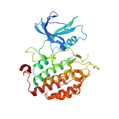
The crystal structure of the catalytic domain of tau tubulin kinase 2 in complex with a small-molecule inhibitor

Marcotte, D.J.Spilker, K.A.Wen, D.Hesson, T.Patterson, T.A.Kumar, R.Chodaparambil, J.V.

(2020) Acta Crystallogr F Struct Biol Commun 76: 103-108

Macromolecule Content 

  • Total Structure Weight: 70.45 kDa 
  • Atom Count: 5,117 
  • Modeled Residue Count: 582 
  • Deposited Residue Count: 600 
  • Unique protein chains: 1

Macromolecules

Find similar proteins by:|  3D Structure
Entity ID: 1
MoleculeChains  Sequence LengthOrganismDetailsImage
Tau-tubulin kinase 2A [auth B],
B [auth A]
300Homo sapiensMutation(s): 0 
Gene Names: TTBK2KIAA0847
EC: 2.7.11.1
UniProt & NIH Common Fund Data Resources
Find proteins for Q6IQ55 (Homo sapiens)
Explore Q6IQ55 
Go to UniProtKB:  Q6IQ55
PHAROS:  Q6IQ55
GTEx:  ENSG00000128881 
Entity Groups
Sequence Clusters30% Identity50% Identity70% Identity90% Identity95% Identity100% Identity
UniProt GroupQ6IQ55
Sequence Annotations
Expand
Reference Sequence

Small Molecules

Ligands 3 Unique
IDChains Name / Formula / InChI Key2D Diagram3D Interactions
DTQ
(Subject of Investigation/LOI)

Query on DTQ


Download:Ideal Coordinates CCD File
E [auth B],
I [auth A]
4-[3-HYDROXYANILINO]-6,7-DIMETHOXYQUINAZOLINE
C16 H15 N3 O3
BNDYIYYKEIXHNK-UHFFFAOYSA-N
PO4

Query on PO4


Download:Ideal Coordinates CCD File
C [auth B]
D [auth B]
F [auth B]
H [auth A]
J [auth A]
C [auth B],
D [auth B],
F [auth B],
H [auth A],
J [auth A],
K [auth A],
M [auth A],
N [auth A]
PHOSPHATE ION
O4 P
NBIIXXVUZAFLBC-UHFFFAOYSA-K
GOL

Query on GOL


Download:Ideal Coordinates CCD File
G [auth B],
L [auth A]
GLYCEROL
C3 H8 O3
PEDCQBHIVMGVHV-UHFFFAOYSA-N

Experimental Data & Validation

Experimental Data

  • Method: X-RAY DIFFRACTION
  • Resolution: 1.74 Å
  • R-Value Free:  0.233 (Depositor), 0.235 (DCC) 
  • R-Value Work:  0.191 (Depositor), 0.195 (DCC) 
  • R-Value Observed: 0.193 (Depositor) 
Space Group: P 21 21 21
Unit Cell:
Length ( Å )Angle ( ˚ )
a = 56.23α = 90
b = 114.68β = 90
c = 118.7γ = 90
Software Package:
Software NamePurpose
PHENIXrefinement
SCALAdata scaling
PDB_EXTRACTdata extraction
MOSFLMdata reduction
PHASERphasing

Structure Validation

View Full Validation Report


Ligand Structure Quality Assessment 


Entry History 

Deposition Data

Revision History  (Full details and data files)

  • Version 1.0: 2020-03-11
    Type: Initial release
  • Version 1.1: 2023-10-11
    Changes: Data collection, Database references, Refinement description