6TXE | pdb_00006txe

Crystal structure of tetrameric human wt-SAMHD1 (residues 109-626) with GTP, dATP, dTMPNPP and Mg


Experimental Data Snapshot

  • Method: X-RAY DIFFRACTION
  • Resolution: 3.19 Å
  • R-Value Free: 
    0.252 (Depositor), 0.251 (DCC) 
  • R-Value Work: 
    0.218 (Depositor), 0.219 (DCC) 
  • R-Value Observed: 
    0.220 (Depositor) 

Starting Model: experimental
View more details

wwPDB Validation 3D Report Full Report

Validation slider image for 6TXE

Ligand Structure Quality Assessment 


This is version 1.2 of the entry. See complete history

Literature

Crystal structures of SAMHD1 inhibitor complexes reveal the mechanism of water-mediated dNTP hydrolysis.

Morris, E.R.Caswell, S.J.Kunzelmann, S.Arnold, L.H.Purkiss, A.G.Kelly, G.Taylor, I.A.

(2020) Nat Commun 11: 3165-3165

  • DOI: https://doi.org/10.1038/s41467-020-16983-2
  • Primary Citation Related Structures: 
    6TX0, 6TXA, 6TXC, 6TXE, 6TXF, 6XU1, 6YOM

  • PubMed Abstract: 

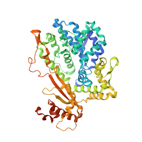
    SAMHD1 regulates cellular 2'-deoxynucleoside-5'-triphosphate (dNTP) homeostasis by catalysing the hydrolysis of dNTPs into 2'-deoxynucleosides and triphosphate. In CD4 + myeloid lineage and resting T-cells, SAMHD1 blocks HIV-1 and other viral infections by depletion of the dNTP pool to a level that cannot support replication. SAMHD1 mutations are associated with the autoimmune disease Aicardi-Goutières syndrome and hypermutated cancers. Furthermore, SAMHD1 sensitises cancer cells to nucleoside-analogue anti-cancer therapies and is linked with DNA repair and suppression of the interferon response to cytosolic nucleic acids. Nevertheless, despite its requirement in these processes, the fundamental mechanism of SAMHD1-catalysed dNTP hydrolysis remained unknown. Here, we present structural and enzymological data showing that SAMHD1 utilises an active site, bi-metallic iron-magnesium centre that positions a hydroxide nucleophile in-line with the P α -O 5' bond to catalyse phosphoester bond hydrolysis. This precise molecular mechanism for SAMHD1 catalysis, reveals how SAMHD1 down-regulates cellular dNTP and modulates the efficacy of nucleoside-based anti-cancer and anti-viral therapies.


  • Organizational Affiliation
    • Macromolecular Structure Laboratory, The Francis Crick Institute, 1 Midland Road, London, NW1 1AT, UK.

Macromolecule Content 

  • Total Structure Weight: 990 kDa 
  • Atom Count: 62,675 
  • Modeled Residue Count: 7,654 
  • Deposited Residue Count: 8,320 
  • Unique protein chains: 1

Macromolecules

Find similar proteins by:|  3D Structure
Entity ID: 1
MoleculeChains  Sequence LengthOrganismDetailsImage
Deoxynucleoside triphosphate triphosphohydrolase SAMHD1
A, B, C, D, E
A, B, C, D, E, F, G, H, I, J, K, L, M, N, O, P
520Homo sapiensMutation(s): 0 
Gene Names: SAMHD1MOP5
EC: 3.1.5
UniProt & NIH Common Fund Data Resources
Find proteins for Q9Y3Z3 (Homo sapiens)
Explore Q9Y3Z3 
Go to UniProtKB:  Q9Y3Z3
PHAROS:  Q9Y3Z3
GTEx:  ENSG00000101347 
Entity Groups
Sequence Clusters30% Identity50% Identity70% Identity90% Identity95% Identity100% Identity
UniProt GroupQ9Y3Z3
Sequence Annotations
Expand
Reference Sequence

Small Molecules

Ligands 6 Unique
IDChains Name / Formula / InChI Key2D Diagram3D Interactions
GTP
(Subject of Investigation/LOI)

Query on GTP


Download:Ideal Coordinates CCD File
AA [auth B]
CB [auth F]
FD [auth N]
KA [auth D]
KB [auth G]
AA [auth B],
CB [auth F],
FD [auth N],
KA [auth D],
KB [auth G],
LC [auth K],
MD [auth O],
PA [auth D],
QB [auth H],
SC [auth L],
SD [auth P],
U [auth A],
VA [auth E],
WB [auth I],
XC [auth M],
YB [auth I]
GUANOSINE-5'-TRIPHOSPHATE
C10 H16 N5 O14 P3
XKMLYUALXHKNFT-UUOKFMHZSA-N
DTP
(Subject of Investigation/LOI)

Query on DTP


Download:Ideal Coordinates CCD File
AD [auth N]
CA [auth B]
DA [auth C]
EB [auth F]
FB [auth G]
AD [auth N],
CA [auth B],
DA [auth C],
EB [auth F],
FB [auth G],
FC [auth J],
GC [auth K],
HD [auth N],
JA [auth D],
LB [auth H],
NC [auth L],
V [auth A],
XA [auth E],
YC [auth M],
ZB [auth I],
ZC [auth M]
2'-DEOXYADENOSINE 5'-TRIPHOSPHATE
C10 H16 N5 O12 P3
SUYVUBYJARFZHO-RRKCRQDMSA-N
1FZ
(Subject of Investigation/LOI)

Query on 1FZ


Download:Ideal Coordinates CCD File
BB [auth F]
DC [auth J]
ED [auth N]
HA [auth C]
JB [auth G]
BB [auth F],
DC [auth J],
ED [auth N],
HA [auth C],
JB [auth G],
KC [auth K],
LD [auth O],
OA [auth D],
PB [auth H],
RC [auth L],
RD [auth P],
T [auth A],
UA [auth E],
VB [auth I],
WC [auth M],
Z [auth B]
5'-O-[(R)-hydroxy{[(R)-hydroxy(phosphonooxy)phosphoryl]amino}phosphoryl]thymidine
C10 H18 N3 O13 P3
YRKUYVYMPXAOAT-XLPZGREQSA-N
SO4

Query on SO4


Download:Ideal Coordinates CCD File
BA [auth B]
DB [auth F]
EC [auth J]
GD [auth N]
IA [auth C]
BA [auth B],
DB [auth F],
EC [auth J],
GD [auth N],
IA [auth C],
MC [auth K],
ND [auth O],
QA [auth D],
RB [auth H],
WA [auth E],
XB [auth I]
SULFATE ION
O4 S
QAOWNCQODCNURD-UHFFFAOYSA-L
FE
(Subject of Investigation/LOI)

Query on FE


Download:Ideal Coordinates CCD File
AC [auth J]
BD [auth N]
EA [auth C]
GB [auth G]
HC [auth K]
AC [auth J],
BD [auth N],
EA [auth C],
GB [auth G],
HC [auth K],
ID [auth O],
LA [auth D],
MB [auth H],
OC [auth L],
OD [auth P],
Q [auth A],
RA [auth E],
SB [auth I],
TC [auth M],
W [auth B],
YA [auth F]
FE (III) ION
Fe
VTLYFUHAOXGGBS-UHFFFAOYSA-N
MG
(Subject of Investigation/LOI)

Query on MG


Download:Ideal Coordinates CCD File
AB [auth F]
BC [auth J]
CC [auth J]
CD [auth N]
DD [auth N]
AB [auth F],
BC [auth J],
CC [auth J],
CD [auth N],
DD [auth N],
FA [auth C],
GA [auth C],
HB [auth G],
IB [auth G],
IC [auth K],
JC [auth K],
JD [auth O],
KD [auth O],
MA [auth D],
NA [auth D],
NB [auth H],
OB [auth H],
PC [auth L],
PD [auth P],
QC [auth L],
QD [auth P],
R [auth A],
S [auth A],
SA [auth E],
TA [auth E],
TB [auth I],
UB [auth I],
UC [auth M],
VC [auth M],
X [auth B],
Y [auth B],
ZA [auth F]
MAGNESIUM ION
Mg
JLVVSXFLKOJNIY-UHFFFAOYSA-N

Experimental Data & Validation

Experimental Data

  • Method: X-RAY DIFFRACTION
  • Resolution: 3.19 Å
  • R-Value Free:  0.252 (Depositor), 0.251 (DCC) 
  • R-Value Work:  0.218 (Depositor), 0.219 (DCC) 
  • R-Value Observed: 0.220 (Depositor) 
Space Group: P 1 21 1
Unit Cell:
Length ( Å )Angle ( ˚ )
a = 97.482α = 90
b = 172.232β = 95.31
c = 275.271γ = 90
Software Package:
Software NamePurpose
XDSdata reduction
Aimlessdata scaling
STARANISOdata scaling
PHASERphasing
REFMACrefinement
PDB_EXTRACTdata extraction

Structure Validation

View Full Validation Report


Ligand Structure Quality Assessment 


Entry History 

& Funding Information

Deposition Data


Funding OrganizationLocationGrant Number
Wellcome TrustUnited Kingdom108014/Z/15/Z
Wellcome TrustUnited KingdomFC001029
Medical Research Council (MRC, United Kingdom)United KingdomFC001029
Cancer Research UKUnited KingdomFC001029

Revision History  (Full details and data files)

  • Version 1.0: 2020-06-24
    Type: Initial release
  • Version 1.1: 2020-07-08
    Changes: Database references
  • Version 1.2: 2024-01-24
    Changes: Data collection, Database references, Derived calculations, Other, Refinement description