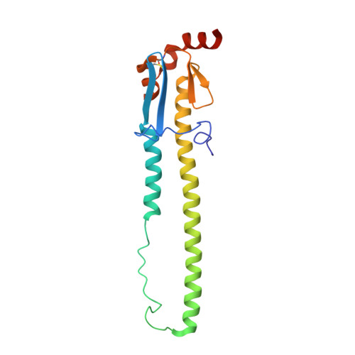
6TWS

Crystal structure of the haemagglutinin mutant (Gln226Leu, Gly228Ser) from an H10N7 seal influenza virus isolated in Germany with 2mM EDTA


Experimental Data Snapshot

  • Method: X-RAY DIFFRACTION
  • Resolution: 2.00 Å
  • R-Value Free: 0.231 
  • R-Value Work: 0.209 
  • R-Value Observed: 0.210 

Starting Model: experimental
View more details

wwPDB Validation   3D Report Full Report


Ligand Structure Quality Assessment 


This is version 1.2 of the entry. See complete history


Literature

Hemagglutinin Traits Determine Transmission of Avian A/H10N7 Influenza Virus between Mammals.

Herfst, S.Zhang, J.Richard, M.McBride, R.Lexmond, P.Bestebroer, T.M.Spronken, M.I.J.de Meulder, D.van den Brand, J.M.Rosu, M.E.Martin, S.R.Gamblin, S.J.Xiong, X.Peng, W.Bodewes, R.van der Vries, E.Osterhaus, A.D.M.E.Paulson, J.C.Skehel, J.J.Fouchier, R.A.M.

(2020) Cell Host Microbe 28: 602-613.e7

  • DOI: https://doi.org/10.1016/j.chom.2020.08.011
  • Primary Citation of Related Structures:  
    6TJW, 6TJY, 6TVA, 6TVB, 6TVC, 6TVD, 6TVF, 6TVR, 6TVS, 6TVT, 6TWH, 6TWI, 6TWS, 6TWV, 6TXO, 6TY1

  • PubMed Abstract: 

    In 2014, an outbreak of avian A/H10N7 influenza virus occurred among seals along North-European coastal waters, significantly impacting seal populations. Here, we examine the cross-species transmission and mammalian adaptation of this influenza A virus, revealing changes in the hemagglutinin surface protein that increase stability and receptor binding. The seal A/H10N7 virus was aerosol or respiratory droplet transmissible between ferrets. Compared with avian H10 hemagglutinin, seal H10 hemagglutinin showed stronger binding to the human-type sialic acid receptor, with preferential binding to α2,6-linked sialic acids on long extended branches. In X-ray structures, changes in the 220-loop of the receptor-binding pocket caused similar interactions with human receptor as seen for pandemic strains. Two substitutions made seal H10 hemagglutinin more stable than avian H10 hemagglutinin and similar to human hemagglutinin. Consequently, identification of avian-origin influenza viruses across mammals appears critical to detect influenza A viruses posing a major threat to humans and other mammals.


  • Organizational Affiliation

    Department of Viroscience, Postgraduate School of Molecular Medicine, Erasmus MC University Medical Center, 3015GE, Rotterdam, the Netherlands.


Macromolecules
Find similar proteins by:  (by identity cutoff)  |  3D Structure
Entity ID: 1
MoleculeChains Sequence LengthOrganismDetailsImage
HemagglutininA [auth G],
C [auth A],
E [auth C]
325Influenza A virus (A/harbour seal/Germany/1/2014(H10N7))Mutation(s): 1 
Gene Names: HA
UniProt
Find proteins for A0A0A7HR51 (Influenza A virus)
Explore A0A0A7HR51 
Go to UniProtKB:  A0A0A7HR51
Entity Groups  
Sequence Clusters30% Identity50% Identity70% Identity90% Identity95% Identity100% Identity
UniProt GroupA0A0A7HR51
Glycosylation
Glycosylation Sites: 1
Sequence Annotations
Expand
  • Reference Sequence
Find similar proteins by:  (by identity cutoff)  |  3D Structure
Entity ID: 2
MoleculeChains Sequence LengthOrganismDetailsImage
Hemagglutinin HA2B [auth H],
D [auth B],
F [auth D]
177Influenza A virus (A/harbour seal/Germany/1/2014(H10N7))Mutation(s): 0 
Gene Names: HA
UniProt
Find proteins for A0A0A7HR51 (Influenza A virus)
Explore A0A0A7HR51 
Go to UniProtKB:  A0A0A7HR51
Entity Groups  
Sequence Clusters30% Identity50% Identity70% Identity90% Identity95% Identity100% Identity
UniProt GroupA0A0A7HR51
Glycosylation
Glycosylation Sites: 1
Sequence Annotations
Expand
  • Reference Sequence
Oligosaccharides

Help

Entity ID: 3
MoleculeChains Length2D Diagram Glycosylation3D Interactions
2-acetamido-2-deoxy-beta-D-glucopyranose-(1-4)-2-acetamido-2-deoxy-beta-D-glucopyranoseG [auth E],
H [auth F],
I
2N-Glycosylation
Glycosylation Resources
GlyTouCan:  G42666HT
GlyCosmos:  G42666HT
GlyGen:  G42666HT
Small Molecules
Ligands 2 Unique
IDChains Name / Formula / InChI Key2D Diagram3D Interactions
NAG
Query on NAG

Download Ideal Coordinates CCD File 
U [auth B],
V [auth C]
2-acetamido-2-deoxy-beta-D-glucopyranose
C8 H15 N O6
OVRNDRQMDRJTHS-FMDGEEDCSA-N
EDO
Query on EDO

Download Ideal Coordinates CCD File 
AA [auth C]
BA [auth C]
CA [auth C]
DA [auth C]
EA [auth C]
AA [auth C],
BA [auth C],
CA [auth C],
DA [auth C],
EA [auth C],
FA [auth D],
GA [auth D],
J [auth G],
K [auth G],
L [auth G],
M [auth H],
N [auth A],
O [auth A],
P [auth A],
Q [auth A],
R [auth A],
S [auth B],
T [auth B],
W [auth C],
X [auth C],
Y [auth C],
Z [auth C]
1,2-ETHANEDIOL
C2 H6 O2
LYCAIKOWRPUZTN-UHFFFAOYSA-N
Experimental Data & Validation

Experimental Data

  • Method: X-RAY DIFFRACTION
  • Resolution: 2.00 Å
  • R-Value Free: 0.231 
  • R-Value Work: 0.209 
  • R-Value Observed: 0.210 
  • Space Group: P 1 21 1
Unit Cell:
Length ( Å )Angle ( ˚ )
a = 65.148α = 90
b = 215.596β = 100.46
c = 72.87γ = 90
Software Package:
Software NamePurpose
REFMACrefinement
PDB_EXTRACTdata extraction
xia2data reduction
Aimlessdata scaling
PHASERphasing

Structure Validation

View Full Validation Report


Ligand Structure Quality Assessment 


Entry History & Funding Information

Deposition Data


Funding OrganizationLocationGrant Number
The Francis Crick InstituteUnited Kingdom--

Revision History  (Full details and data files)

  • Version 1.0: 2020-10-21
    Type: Initial release
  • Version 1.1: 2024-01-24
    Changes: Data collection, Database references, Refinement description
  • Version 1.2: 2024-11-06
    Changes: Structure summary