6TWM | pdb_00006twm

Product bound structure of the Ectoine utilization protein EutE (DoeB) from Ruegeria pomeroyi


Experimental Data Snapshot

  • Method: X-RAY DIFFRACTION
  • Resolution: 2.50 Å
  • R-Value Free: 
    0.262 (Depositor), 0.259 (DCC) 
  • R-Value Work: 
    0.209 (Depositor), 0.207 (DCC) 
  • R-Value Observed: 
    0.209 (Depositor) 

Starting Model: experimental
View more details

wwPDB Validation 3D Report Full Report

Validation slider image for 6TWM

Ligand Structure Quality Assessment 


This is version 2.0 of the entry. See complete history

Literature

Degradation of the microbial stress protectants and chemical chaperones ectoine and hydroxyectoine by a bacterial hydrolase-deacetylase complex.

Mais, C.N.Hermann, L.Altegoer, F.Seubert, A.Richter, A.A.Wernersbach, I.Czech, L.Bremer, E.Bange, G.

(2020) J Biological Chem 295: 9087-9104

  • DOI: https://doi.org/10.1074/jbc.RA120.012722
  • Primary Citation Related Structures: 
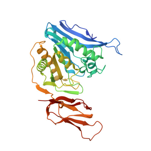
    6TWJ, 6TWK, 6TWL, 6TWM, 6YO9

  • PubMed Abstract: 

    When faced with increased osmolarity in the environment, many bacterial cells accumulate the compatible solute ectoine and its derivative 5-hydroxyectoine. Both compounds are not only potent osmostress protectants, but also serve as effective chemical chaperones stabilizing protein functionality. Ectoines are energy-rich nitrogen and carbon sources that have an ecological impact that shapes microbial communities. Although the biochemistry of ectoine and 5-hydroxyectoine biosynthesis is well understood, our understanding of their catabolism is only rudimentary. Here, we combined biochemical and structural approaches to unravel the core of ectoine and 5-hydroxy-ectoine catabolisms. We show that a conserved enzyme bimodule consisting of the EutD ectoine/5-hydroxyectoine hydrolase and the EutE deacetylase degrades both ectoines. We determined the high-resolution crystal structures of both enzymes, derived from the salt-tolerant bacteria Ruegeria pomeroyi and Halomonas elongata These structures, either in their apo-forms or in forms capturing substrates or intermediates, provided detailed insights into the catalytic cores of the EutD and EutE enzymes. The combined biochemical and structural results indicate that the EutD homodimer opens the pyrimidine ring of ectoine through an unusual covalent intermediate, N -α-2 acetyl-l-2,4-diaminobutyrate (α-ADABA). We found that α-ADABA is then deacetylated by the zinc-dependent EutE monomer into diaminobutyric acid (DABA), which is further catabolized to l-aspartate. We observed that the EutD-EutE bimodule synthesizes exclusively the α-, but not the γ-isomers of ADABA or hydroxy-ADABA. Of note, α-ADABA is known to induce the MocR/GabR-type repressor EnuR, which controls the expression of many ectoine catabolic genes clusters. We conclude that hydroxy-α-ADABA might serve a similar function.


  • Organizational Affiliation
    • Philipps-University Marburg, Center for Synthetic Microbiology (SYNMIKRO) & Faculty of Chemistry, Marburg, Germany.

Macromolecule Content 

  • Total Structure Weight: 434.22 kDa 
  • Atom Count: 30,865 
  • Modeled Residue Count: 3,916 
  • Deposited Residue Count: 4,044 
  • Unique protein chains: 1

Macromolecules

Find similar proteins by:|  3D Structure
Entity ID: 1
MoleculeChains  Sequence LengthOrganismDetailsImage
N-acetyl-L-2,4-diaminobutyric acid deacetylase
A, B, C, D, E
A, B, C, D, E, F, G, H, I, J, K, L
337Ruegeria pomeroyi DSS-3Mutation(s): 0 
Gene Names: doeBSPO1139
UniProt
Find proteins for Q5LUB5 (Ruegeria pomeroyi (strain ATCC 700808 / DSM 15171 / DSS-3))
Explore Q5LUB5 
Go to UniProtKB:  Q5LUB5
Entity Groups
Sequence Clusters30% Identity50% Identity70% Identity90% Identity95% Identity100% Identity
UniProt GroupQ5LUB5
Sequence Annotations
Expand
Reference Sequence

Small Molecules

Ligands 3 Unique
IDChains Name / Formula / InChI Key2D Diagram3D Interactions
DAB
(Subject of Investigation/LOI)

Query on DAB


Download:Ideal Coordinates CCD File
DA [auth J]
EA [auth K]
N [auth A]
P [auth B]
T [auth D]
DA [auth J],
EA [auth K],
N [auth A],
P [auth B],
T [auth D],
V [auth E],
W [auth F]
2,4-DIAMINOBUTYRIC ACID
C4 H10 N2 O2
OGNSCSPNOLGXSM-VKHMYHEASA-N
ZN

Query on ZN


Download:Ideal Coordinates CCD File
BA [auth I]
CA [auth J]
FA [auth L]
M [auth A]
O [auth B]
BA [auth I],
CA [auth J],
FA [auth L],
M [auth A],
O [auth B],
Q [auth C],
S [auth D],
U [auth E],
X [auth G],
Z [auth H]
ZINC ION
Zn
PTFCDOFLOPIGGS-UHFFFAOYSA-N
ACT

Query on ACT


Download:Ideal Coordinates CCD File
AA [auth H],
R [auth C],
Y [auth G]
ACETATE ION
C2 H3 O2
QTBSBXVTEAMEQO-UHFFFAOYSA-M

Experimental Data & Validation

Experimental Data

  • Method: X-RAY DIFFRACTION
  • Resolution: 2.50 Å
  • R-Value Free:  0.262 (Depositor), 0.259 (DCC) 
  • R-Value Work:  0.209 (Depositor), 0.207 (DCC) 
  • R-Value Observed: 0.209 (Depositor) 
Space Group: P 1 21 1
Unit Cell:
Length ( Å )Angle ( ˚ )
a = 76.667α = 90
b = 145.949β = 92.301
c = 164.116γ = 90
Software Package:
Software NamePurpose
PHENIXrefinement
Cootmodel building
XDSdata reduction
XSCALEdata scaling
PHASERphasing

Structure Validation

View Full Validation Report


Ligand Structure Quality Assessment 


Entry History 

Deposition Data

Revision History  (Full details and data files)

  • Version 1.0: 2020-05-20
    Type: Initial release
  • Version 1.1: 2020-05-27
    Changes: Database references
  • Version 1.2: 2020-07-15
    Changes: Database references
  • Version 1.3: 2024-01-24
    Changes: Data collection, Database references, Derived calculations, Refinement description
  • Version 2.0: 2025-10-01
    Changes: Advisory, Atomic model, Data collection, Derived calculations, Structure summary