


Crystal structure of the GGDEF domain of DgcB from Caulobacter crescentus in complex with c-di-GMP
Holzschuh, F., Schirmer, T., Teixeira, R.To be published.
Experimental Data Snapshot
Starting Models: experimental
View more details
Entity ID: 1 | |||||
|---|---|---|---|---|---|
| Molecule | Chains | Sequence Length | Organism | Details | Image |
| GGDEF diguanylate cyclase DgcB | 199 | Caulobacter vibrioides NA1000 | Mutation(s): 0 Gene Names: dgcB, CCNA_01926 EC: 2.7.7.65 | 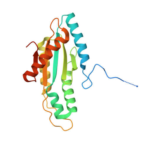 | |
UniProt | |||||
Find proteins for A0A0H3CAN8 (Caulobacter vibrioides (strain NA1000 / CB15N)) Explore A0A0H3CAN8 Go to UniProtKB: A0A0H3CAN8 | |||||
Entity Groups | |||||
| Sequence Clusters | 30% Identity50% Identity70% Identity90% Identity95% Identity100% Identity | ||||
| UniProt Group | A0A0H3CAN8 | ||||
Sequence AnnotationsExpand | |||||
Reference Sequence | |||||
| Ligands 2 Unique | |||||
|---|---|---|---|---|---|
| ID | Chains | Name / Formula / InChI Key | 2D Diagram | 3D Interactions | |
| C2E Download:Ideal Coordinates CCD File | C [auth A], G [auth A], H [auth A], I [auth A], M [auth B] | 9,9'-[(2R,3R,3aS,5S,7aR,9R,10R,10aS,12S,14aR)-3,5,10,12-tetrahydroxy-5,12-dioxidooctahydro-2H,7H-difuro[3,2-d:3',2'-j][1,3,7,9,2,8]tetraoxadiphosphacyclododecine-2,9-diyl]bis(2-amino-1,9-dihydro-6H-purin-6-one) C20 H24 N10 O14 P2 PKFDLKSEZWEFGL-MHARETSRSA-N |  | ||
| SO4 Download:Ideal Coordinates CCD File | D [auth A] E [auth A] F [auth A] J [auth B] K [auth B] | SULFATE ION O4 S QAOWNCQODCNURD-UHFFFAOYSA-L |  | ||
| Length ( Å ) | Angle ( ˚ ) |
|---|---|
| a = 64.5 | α = 90 |
| b = 64.5 | β = 90 |
| c = 246.76 | γ = 90 |
| Software Name | Purpose |
|---|---|
| PHENIX | refinement |
| iMOSFLM | data reduction |
| Aimless | data scaling |
| PHASER | phasing |
| Funding Organization | Location | Grant Number |
|---|---|---|
| Swiss National Science Foundation | Switzerland | 31003A-166652 |


