6TT0 | pdb_00006tt0

Crystal structure of a potent and reversible dual binding site Acetylcholinesterase chiral inhibitor


Experimental Data Snapshot

  • Method: X-RAY DIFFRACTION
  • Resolution: 2.80 Å
  • R-Value Free: 
    0.235 (Depositor), 0.237 (DCC) 
  • R-Value Work: 
    0.188 (Depositor), 0.193 (DCC) 
  • R-Value Observed: 
    0.191 (Depositor) 

Starting Model: experimental
View more details

wwPDB Validation 3D Report Full Report

Validation slider image for 6TT0

Ligand Structure Quality Assessment 


This is version 1.5 of the entry. See complete history

Literature

Chiral Separation, X-ray Structure, and Biological Evaluation of a Potent and Reversible Dual Binding Site AChE Inhibitor.

Catto, M.Pisani, L.de la Mora, E.Belviso, B.D.Mangiatordi, G.F.Pinto, A.Palma, A.Denora, N.Caliandro, R.Colletier, J.P.Silman, I.Nicolotti, O.Altomare, C.D.

(2020) ACS Med Chem Lett 11: 869-876

  • DOI: https://doi.org/10.1021/acsmedchemlett.9b00656
  • Primary Citation Related Structures: 
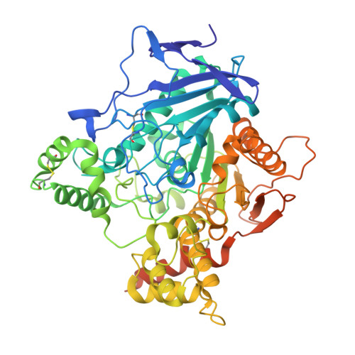
    6TT0

  • PubMed Abstract: 

    Acetylcholinesterase (AChE) inhibitors (AChEIs) still remain the leading therapeutic options for the symptomatic treatment of cognitive deficits associated with mild-to-moderate Alzheimer's disease. The search for new AChEIs benefits from well-established knowledge of the molecular interactions of selective AChEIs, such as donepezil and related dual binding site inhibitors. Starting from a previously disclosed coumarin-based inhibitor (±)- cis - 1 , active as racemate in the nanomolar range toward AChE, we proceeded on a double track by (i) achieving chiral resolution of the enantiomers of 1 by HPLC and (ii) preparing two close achiral analogues of 1 , i.e., compounds 4 and 6 . An eudismic ratio as high as 20 was observed for the (-) enantiomer of cis - 1 . The X-ray crystal structure of the complex between the (-)- cis - 1 eutomer (coded as MC1420 ) and T. californica AChE was determined at 2.8 Å, and docking calculation results suggested that the eutomer in (1 R ,3 S ) absolute configuration should be energetically more favored in binding the enzyme than the eutomer in (1 S ,3 R ) configuration. The achiral analogues 4 and 6 were less effective in inhibiting AChE compared to (±)- cis - 1 , but interestingly butylamide 4 emerged as a potent inhibitor of butyrylcholinesterase (BChE).


  • Organizational Affiliation
    • Department of Pharmacy-Drug Sciences, University of Bari Aldo Moro, Via E. Orabona 4, 70125 Bari, Italy.

Macromolecule Content 

  • Total Structure Weight: 66.19 kDa 
  • Atom Count: 4,392 
  • Modeled Residue Count: 532 
  • Deposited Residue Count: 572 
  • Unique protein chains: 1

Macromolecules

Find similar proteins by:|  3D Structure
Entity ID: 1
MoleculeChains  Sequence LengthOrganismDetailsImage
Acetylcholinesterase572Tetronarce californicaMutation(s): 0 
Gene Names: ache
EC: 3.1.1.7
UniProt
Find proteins for P04058 (Tetronarce californica)
Explore P04058 
Go to UniProtKB:  P04058
Entity Groups
Sequence Clusters30% Identity50% Identity70% Identity90% Identity95% Identity100% Identity
UniProt GroupP04058
Glycosylation
Glycosylation Sites: 1
Sequence Annotations
Expand
Reference Sequence

Small Molecules

Ligands 2 Unique
IDChains Name / Formula / InChI Key2D Diagram3D Interactions
N9T
(Subject of Investigation/LOI)

Query on N9T


Download:Ideal Coordinates CCD File
B [auth A](1~{R},3~{S})-~{N}-(6,7-dimethoxy-2-oxidanylidene-chromen-3-yl)-3-[(phenylmethyl)amino]cyclohexane-1-carboxamide
C25 H28 N2 O5
UGTGSTDFYWUXNH-MJGOQNOKSA-N
NAG

Query on NAG


Download:Ideal Coordinates CCD File
C [auth A],
D [auth A]
2-acetamido-2-deoxy-beta-D-glucopyranose
C8 H15 N O6
OVRNDRQMDRJTHS-FMDGEEDCSA-N

Experimental Data & Validation

Experimental Data

  • Method: X-RAY DIFFRACTION
  • Resolution: 2.80 Å
  • R-Value Free:  0.235 (Depositor), 0.237 (DCC) 
  • R-Value Work:  0.188 (Depositor), 0.193 (DCC) 
  • R-Value Observed: 0.191 (Depositor) 
Space Group: P 31 2 1
Unit Cell:
Length ( Å )Angle ( ˚ )
a = 111.42α = 90
b = 111.42β = 90
c = 137.37γ = 120
Software Package:
Software NamePurpose
PHENIXrefinement
PHENIXrefinement
XDSdata reduction
XSCALEdata scaling
PHENIXphasing

Structure Validation

View Full Validation Report


Ligand Structure Quality Assessment 


Entry History 

Deposition Data

Revision History  (Full details and data files)

  • Version 1.0: 2020-06-10
    Type: Initial release
  • Version 1.1: 2020-07-29
    Type: Remediation
    Reason: Carbohydrate remediation
    Changes: Data collection, Derived calculations, Structure summary
  • Version 1.2: 2021-05-12
    Changes: Derived calculations, Structure summary
  • Version 1.3: 2023-10-04
    Changes: Data collection, Database references
  • Version 1.4: 2024-01-24
    Changes: Refinement description
  • Version 1.5: 2024-11-20
    Changes: Structure summary